. 2010 Oct 6:3:252.
doi: 10.1186/1756-0500-3-252.

Cloning and characterization of a novel alternatively spliced transcript of the human CHD7 putative helicase

Affiliations

Cloning and characterization of a novel alternatively spliced transcript of the human CHD7 putative helicase

Christian Colin et al. BMC Res Notes. .

Abstract

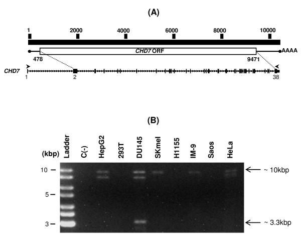
Background: The CHD7 (Chromodomain Helicase DNA binding protein 7) gene encodes a member of the chromodomain family of ATP-dependent chromatin remodeling enzymes. Mutations in the CHD7 gene are found in individuals with CHARGE, a syndrome characterized by multiple birth malformations in several tissues. CHD7 was identified as a binding partner of PBAF complex (Polybromo and BRG Associated Factor containing complex) playing a central role in the transcriptional reprogramming process associated to the formation of multipotent migratory neural crest, a transient cell population associated with the genesis of various tissues. CHD7 is a large gene containing 38 annotated exons and spanning 200 kb of genomic sequence. Although genes containing such number of exons are expected to have several alternative transcripts, there are very few evidences of alternative transcripts associated to CHD7 to date indicating that alternative splicing associated to this gene is poorly characterized.

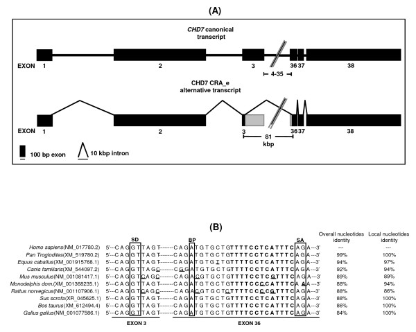
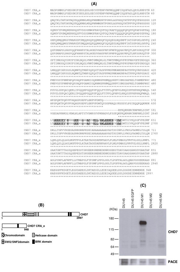
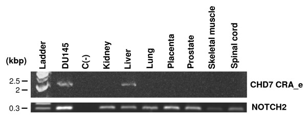
Findings: Here, we report the cloning and characterization by experimental and computational studies of a novel alternative transcript of the human CHD7 (named CHD7 CRA_e), which lacks most of its coding exons. We confirmed by overexpression of CHD7 CRA_e alternative transcript that it is translated into a protein isoform lacking most of the domains displayed by the canonical isoform. Expression of the CHD7 CRA_e transcript was detected in normal liver, in addition to the DU145 human prostate carcinoma cell line from which it was originally isolated.

Conclusions: Our findings indicate that the splicing event associated to the CHD7 CRA_e alternative transcript is functional. The characterization of the CHD7 CRA_e novel isoform presented here not only sets the basis for more detailed functional studies of this isoform, but, also, contributes to the alternative splicing annotation of the CHD7 gene and the design of future functional studies aimed at the elucidation of the molecular functions of its gene products.


Figures

Figure 1
Figure 1
Identification of a putative novel splice variant of CHD7. (A) Representation of the full-length CHD7 transcript structure and primers used to amplify it. At the top the nucleotide sequence numbering scheme of the full-length transcript is represented. At the middle is a schematic representation of the full-length CHD7 transcript with its ORF indicated as a box (the ORF initial and final nucleotides numbers are indicated below the box). At the bottom the exon-intron structure of CHD7 is represented. Exons are represented as black rectangles and intronic sequences as a thin line. The sites of the ORF initial and final nucleotides (dotted lines) and the primers (arrows) annealing to sequences corresponding to exons 1 and 38 of the CHD7 transcript sequence (deposited under the accession number NM_017780.2) used for the amplification of the full-length CHD7 transcript are indicated. (B) Amplification of the full-length CHD7 transcript by RT-PCR. Poly A+ RNA was extracted from the indicated cell lines, and reverse transcription followed by PCR was performed using the CHD7 primers set depicted in (A). Aside from the predicted 10 kbp band, additional RT-PCR products of approximately 9 kbp (not indicated) and 3.3 kbp (indicated) were detected. C(-) is the negative control (PCR reaction without cDNA).
Figure 2
Figure 2
Analysis of the exon-intron structure of the CHD7 CRA_e novel transcript. (A) In the upper part of the figure the exon-intron structure of the canonical CHD7 gene (accession number NM_017780.2) is represented. Exons 4 to 35 have been omitted for clarity. In the lower part the exon-intron structure of the CHD7 CRA_e alternative transcript deduced from sequencing and annealing to the canonical transcript sequence is represented. Splicing is represented as bold lines and numbers below boxes indicate the exon numbers. Rectangles represent the exons, with 0.25 cm in length being equivalent to 100 nucleotides, and the bold line represents the introns, with 0.5 cm being equivalent to 10 kilobase pairs. Regions of alternatively spliced exons 3 and 36 are lightly shaded. (B) Comparative alignment of nucleotide sequences around exonics splicing sites at exons 3 and 36 of human CHD7 with the corresponding exonics regions in Chimpanzee, Horse, Dog, Mouse, short-tailed Opossum, Rat, Boar, Cattle and Chicken. The splicing donor (SD), putative branch point (BP) and splicing acceptor (SA) sites are boxed. The pyrimidine-rich region is shown in bold. Nucleotides differing from the human sequence are underlined.
Figure 3
Figure 3
Deduced protein sequence coded by the CHD7 CRA_e alternative transcript and respective Western blot analysis. (A) Alignment of the putative amino acids sequence coded by the CHD7 CRA_e alternative transcript with the amino acids sequence of the canonical CHD7 protein (accession number: NP_060250). The amino acids in bold and subtitled are conserved in the BRK domain (smart00592). The amino acids from the canonical CHD7 protein that are missing in the CHD7 CRA_e putative amino acid sequence were omitted for clarity (amino acids 601 to 2580). The numbers on the right refer to the amino acids at the end of each line related to each sequence. (B) Schematic representation of the domain structure of canonical CHD7 protein and CHD7 CRA_e isoform. (C) Western blot analysis of DU145 cells stably expressing CHD7 CRA_e alternative transcript (DU145 M1) using an anti-CHD7 antibody which recognizes the C-terminus end of CHD7. Parental DU145 cells and cells transduced with the empty vector (DU145 EGFP) or stably expressing two additional clones of CHD7 CRA_e alternative transcript, which putatively code for truncated proteins (DU145 M2 and DU145 M3) were used as negative controls. Detection of the PACE protein expression is shown as an internal protein loading control.
Figure 4
Figure 4
RT-PCR analysis of the CHD7 CRA_e transcript expression in normal human tissue total RNA samples. To detect the novel CHD7 transcript by RT-PCR we used primers adjacent to the alternative splice site which generate a DNA fragment of approximately 2 kbp. As internal control of the reaction we performed RT-PCR of the same samples using primers specific for the NOTCH2 transcript which yield a DNA fragment of approximately 300 bp (lower part of the figure). A negative control without cDNA was run with each reaction.

References

    1. Vissers LE, van Ravenswaaij CM, Admiraal R, Hurst JA, de Vries BB, Janssen IM, van der Vliet WA, Huys EH, de Jong PJ, Hamel BC, Schoenmakers EF, Brunner HG, Veltman JA, van Kessel AG. Mutations in a new member of the chromodomain gene family cause CHARGE syndrome. Nat Genet. 2004;36:955–957. - PubMed
    1. Sanlaville D, Verloes A. CHARGE syndrome: an update. Eur J Hum Genet. 2007;15:389–399. - PubMed
    1. Aramaki M, Udaka T, Kosaki R, Makita Y, Okamoto N, Yoshihashi H, Oki H, Nanao K, Moriyama N, Oku S, Hasegawa T, Takahashi T, Fukushima Y, Kawame H, Kosaki K. Phenotypic spectrum of CHARGE syndrome with CHD7 mutations. J Pediatr. 2006;148:410–414. - PubMed
    1. Lalani SR, Safiullah AM, Fernbach SD, Harutyunyan KG, Thaller C, Peterson LE, McPherson JD, Gibbs RA, White LD, Hefner M, Davenport SL, Graham JM, Bacino CA, Glass NL, Towbin JA, Craigen WJ, Neish SR, Lin AE, Belmont JW. Spectrum of CHD7 mutations in 110 individuals with CHARGE syndrome and genotype-phenotype correlation. Am J Hum Genet. 2006;78:303–314. - PMC - PubMed
    1. Bosman EA, Penn AC, Ambrose JC, Kettleborough R, Stemple DL, Steel KP. Multiple mutations in mouse Chd7 provide models for CHARGE syndrome. Hum Mol Genet. 2005;14:3463–3476. - PubMed

LinkOut - more resources