Comparative Study
. 2010 Oct 6;11(1):138.
doi: 10.1186/1465-9921-11-138.

Dimethylthiourea protects against chlorine induced changes in airway function in a murine model of irritant induced asthma

Affiliations
Comparative Study

Dimethylthiourea protects against chlorine induced changes in airway function in a murine model of irritant induced asthma

Toby K McGovern et al. Respir Res. .

Abstract

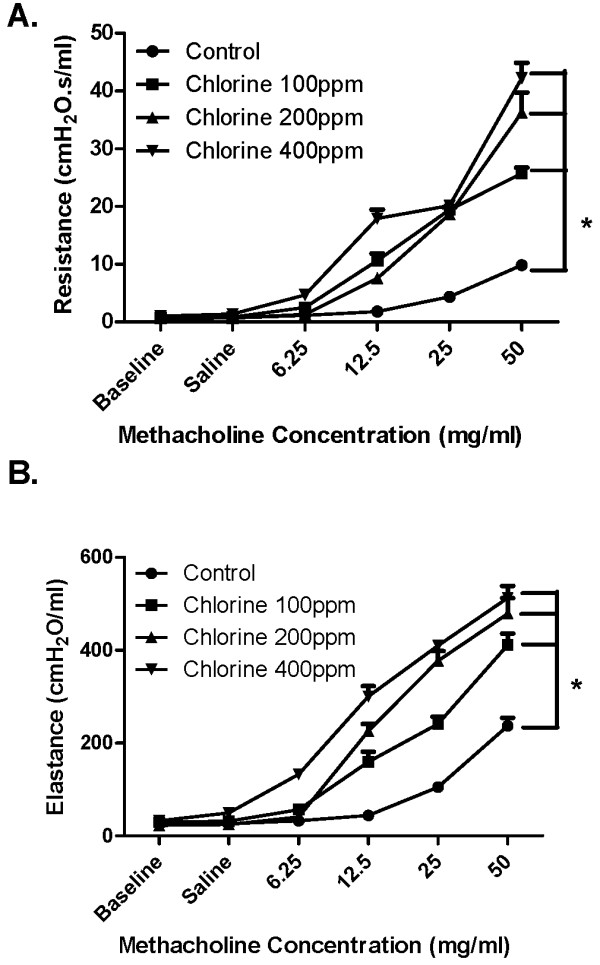
Background: Exposure to chlorine (Cl2) causes airway injury, characterized by oxidative damage, an influx of inflammatory cells and airway hyperresponsiveness. We hypothesized that Cl2-induced airway injury may be attenuated by antioxidant treatment, even after the initial injury.

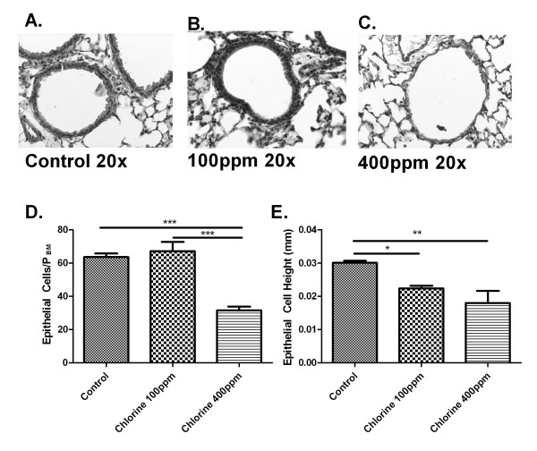
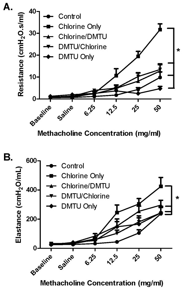
Methods: Balb/C mice were exposed to Cl2 gas (100 ppm) for 5 mins, an exposure that was established to alter airway function with minimal histological disruption of the epithelium. Twenty-four hours after exposure to Cl2, airway responsiveness to aerosolized methacholine (MCh) was measured. Bronchoalveolar lavage (BAL) was performed to determine inflammatory cell profiles, total protein, and glutathione levels. Dimethylthiourea (DMTU;100 mg/kg) was administered one hour before or one hour following Cl2 exposure.

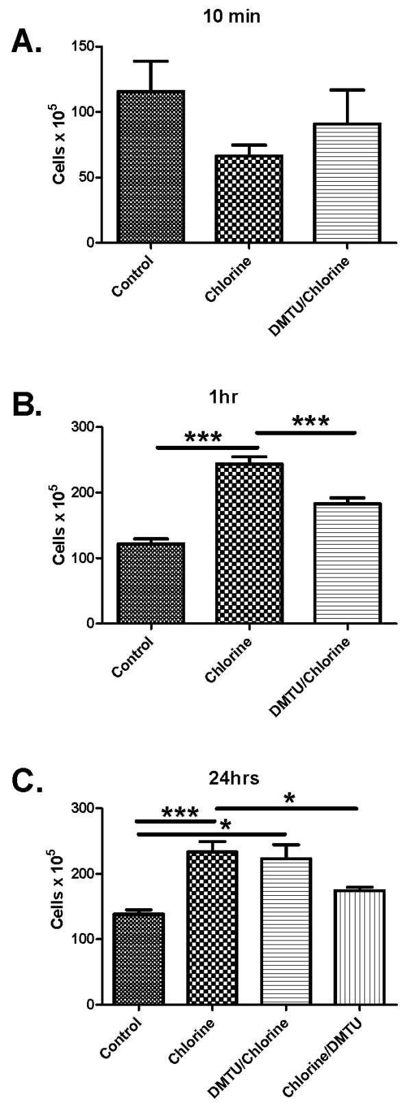
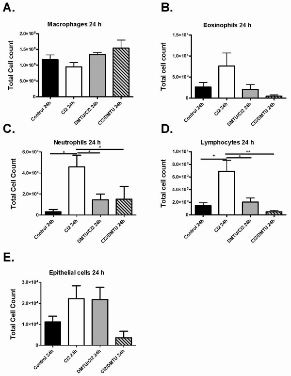
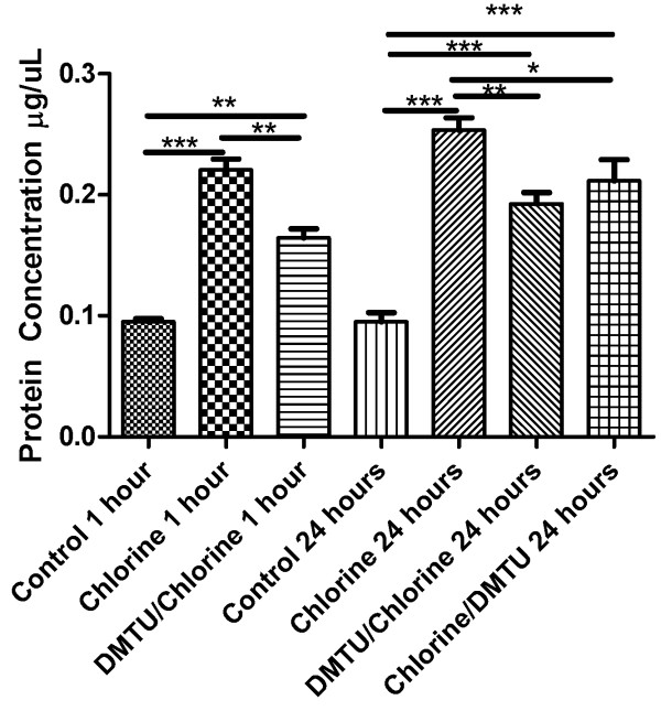
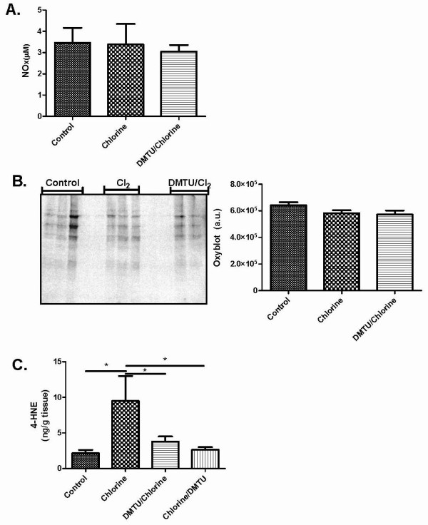
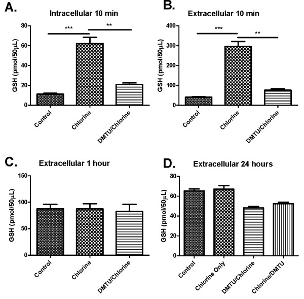
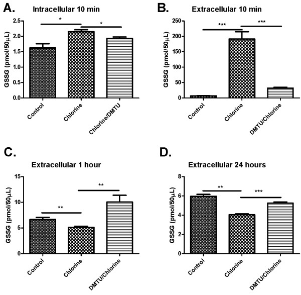
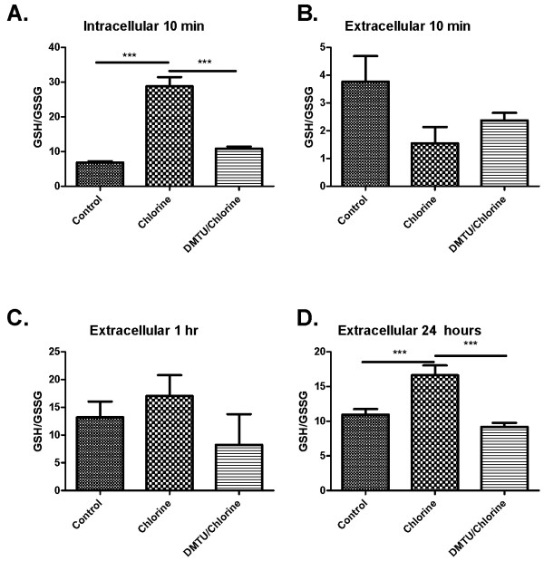
Results: Mice exposed to Cl2 had airway hyperresponsiveness to MCh compared to control animals pre-treated and post-treated with DMTU. Total cell counts in BAL fluid were elevated by Cl2 exposure and were not affected by DMTU treatment. However, DMTU-treated mice had lower protein levels in the BAL than the Cl2-only treated animals. 4-Hydroxynonenal analysis showed that DMTU given pre- or post-Cl2 prevented lipid peroxidation in the lung. Following Cl2 exposure glutathione (GSH) was elevated immediately following exposure both in BAL cells and in fluid and this change was prevented by DMTU. GSSG was depleted in Cl2 exposed mice at later time points. However, the GSH/GSSG ratio remained high in chlorine exposed mice, an effect attenuated by DMTU.

Conclusion: Our data show that the anti-oxidant DMTU is effective in attenuating Cl2 induced increase in airway responsiveness, inflammation and biomarkers of oxidative stress.


Figures

Figure 1
Figure 1
Dose-response effect of Cl2 on respiratory responsiveness to methacholine. Mice were either unchallenged (Control; n = 6) or challenged with 100 (n = 6), 200 (n = 6) or 400 (n = 6) ppm Cl2 gas. After 24 h, total respiratory system resistance (A) and respiratory system elastance (B) in response to saline (Sal) and doubling doses of MCh were assessed using a small animal ventilator (FlexiVent). Baseline (Base) values obtained from untreated mice are shown for comparison. Mice treated with all three concentrations of Cl2 showed significantly higher respiratory system resistance and at 12.5, 25, and 50 mg/ml of MCh as compared with control. * p < 0.05, n = 6 per group.
Figure 2
Figure 2
Effects of Cl2 on airway histology. Twenty-four hours following Cl2 exposure lungs were collected, paraffin embedded and lung sections cut (5 μM). Sections were then stained with hematoxylin and eosin. Representative pictures of airway sections from control mice (A) mice treated with 100 (B), or 400 ppm (C) Cl2. Total epithelial cells were quantified in each airway and corrected for PBM and showed no difference between control and 100 ppm, but significantly fewer epithelial cells at 400 ppm (D). Epithelial cell height was also calculated and showed that mice given 100 ppm and 400 ppm had shorter epithelial cells than control (E).
Figure 3
Figure 3
Effects of Cl2 on methacholine respiratory system resistance and elastance. Panel A shows the effects of Cl2 exposure on total respiratory system resistance in mice that were treated with before and 1 hour after exposure with DMTU. A two-way ANOVA showed that there is a significant difference between mice pre- or post-treated with DMTU when compared to animals receiving Cl2 only. Panel B shows the effects of Cl2 exposure and DMTU treatment on total respiratory system elastance. DMTU/Cl2 treated animals had elastance levels similar to control whereas Cl2 only treated mice had significantly higher values compared to control: n = 6 per group; * p < 0.05.
Figure 4
Figure 4
Effects of Cl2 exposure on the numbers of cells in BAL fluid. Data for control and Cl2 exposed animals that were sacrificed 10 minutes (A), 1 hour (B) and 24 hours (C) after Cl2 exposure. Cl2 exposure caused a significant increase in total leukocytes compared to controls at 1 hour and 24 hours, the effect of which was attenuated by pre-treatment with DMTU at one hour and post treatment with DMTU at 24 hours. (n = 6 per group; * p < 0.05., **p < 0.01, ***p < 0.001).
Figure 5
Figure 5
Cellular composition of BAL fluid following Cl2 exposure. Differential cell counts were done at 10 minutes and 24 hours. No cell subset was significantly different at 10 min (data not shown). At 24 hours neutrophils and lymphocytes were significantly elevated in Cl2 groups. Treatment with DMTU was limited increases in these cell types. There was no difference between control and DMTU treated groups. Control (n = 9), Cl2 100 ppm (n = 7), DMTU/Cl2 (n = 7), Cl2 /DMTU (n = 6); * <0.05.
Figure 6
Figure 6
Effects of Cl2 exposure and DMTU treatment on BAL fluid protein. Protein levels in BAL fluid were assessed by Bradford assay. There was a significant increase in total protein at 1 and 24 hours after Cl2 exposure. Pre-treatment with DMTU attenuated the increase in protein at both time points and at 24 hours when given one hour post- Cl2 exposure. (n = 6-9/group; * p < 0.05, **p < 0.01, ***p < 0.001).
Figure 7
Figure 7
Effects of Cl2 exposure and DMTU treatment on markers of oxidative stress. (A) Nitric oxide was also measured 24 hours following Cl2 exposure using a Griess reaction and no significant change was seen between any of groups. (B) Twenty-four hours following Cl2 exposure BAL was collected and an OxyBlot was performed on lung tissue homogenates to detect carbonylated proteins. No significant differences were detected among the groups. (C) Twenty-four hours following chlorine exposure, lungs were collected for 4-HNE analysis. Chlorine caused a significant increase in 4-HNE levels over control and DMTU treated groups. There were no differences between DMTU groups and baseline. (n = 6-10, * p < 0.05).
Figure 8
Figure 8
Effects of Cl2 exposure and DMTU treatment on glutathione levels in BAL fluid and cells. (A) 10 minutes following Cl2 exposure, GSH levels in the BAL cell fraction show a significant increase that was attenuated by pre-treating the mice with DMTU one hour prior to Cl2 challenge. (B) 10 minutes following Cl2 challenge, the same significant increase of GSH is seen in the BAL supernatant. (C) GSH levels 1 hour following Cl2 exposure and (D) 24 hours after Cl2 exposure were not different among groups. (n = 6-9; * p < 0.05, **p < 0.01, ***p < 0.001).
Figure 9
Figure 9
Effects of Cl2 exposure and DMTU treatment on oxidized glutathione in BAL fluid cells and supernatant. Ten minutes after Cl2 exposure (A-B), oxidized GSSG levels were determined. Animals exposed to Cl2 had increased GSSG in the BAL fluid and intracellularly 10 min following Cl2 exposure (A & B). Extracellular GSSG was reduced at one hour and 24 hours following Cl2 challenge, but no differences were found between control and DMTU treated groups.(n = 6-9; * p < 0.05, **p < 0.01, ***p < 0.001).
Figure 10
Figure 10
Effect of Cl2 exposure and DMTU on ratio of GSH/GSSG. (A) Ten minutes following Cl2 exposure the ratio of GSH/GSSG in the intracellular fraction of the BAL was significantly increased in Cl2 exposed mice compared to control and DMTU/Cl2 treated animals. (B-C) The extracellular fractions of the BAL at ten minutes and 1 hour showed no differences between groups. (D) Cl2 exposure induced a significant increase in the ratio of GSH/GSSG, an effect attenuated by DMTU. (n = 6-9; * p < 0.05, **p < 0.01, ***p < 0.001).

References

    1. Henneberger PK, Olin AC, Andersson E, Hagberg S, Toren K. The incidence of respiratory symptoms and diseases among pulp mill workers with peak exposures to ozone and other irritant gases. Chest. 2005;128:3028. doi: 10.1378/chest.128.4.3028. - DOI - PubMed
    1. Brooks SM, Weiss MA, Bernstein IL. Reactive airways dysfunction syndrome (RADS). Persistent asthma syndrome after high level irritant exposures. Chest. 1985;88:376. doi: 10.1378/chest.88.3.376. - DOI - PubMed
    1. Deschamps D, Soler P, Rosenberg N, Baud F, Gervais P. Persistent Asthma After Inhalation of A Mixture of Sodium-Hypochlorite and Hydrochloric-Acid. Chest. 1994;105:1895. doi: 10.1378/chest.105.6.1895. - DOI - PubMed
    1. Van SD, Wenck MA, Belflower A, Drociuk D, Ferdinands J, Holguin F, Svendsen E, Bretous L, Jankelevich S, Gibson JJ, Garbe P, Moolenaar RL. Acute health effects after exposure to chlorine gas released after a train derailment. Am J Emerg Med. 2009;27:1. doi: 10.1016/j.ajem.2007.12.006. - DOI - PMC - PubMed
    1. Lemiere C, Malo JL, Boutet M. Reactive airways dysfunction syndrome due to chlorine: sequential bronchial biopsies and functional assessment. Eur Respir J. 1997;10:241. doi: 10.1183/09031936.97.10010241. - DOI - PubMed

Publication types