. 2010 Nov 15;70(22):9434-43.
doi: 10.1158/0008-5472.CAN-10-1960. Epub 2010 Oct 5.

Clld7, a candidate tumor suppressor on chromosome 13q14, regulates pathways of DNA damage/repair and apoptosis

Affiliations

Clld7, a candidate tumor suppressor on chromosome 13q14, regulates pathways of DNA damage/repair and apoptosis

Xiaobo Zhou et al. Cancer Res. .

Abstract

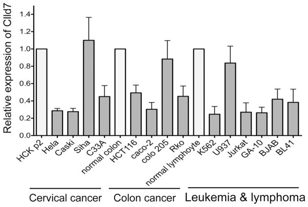
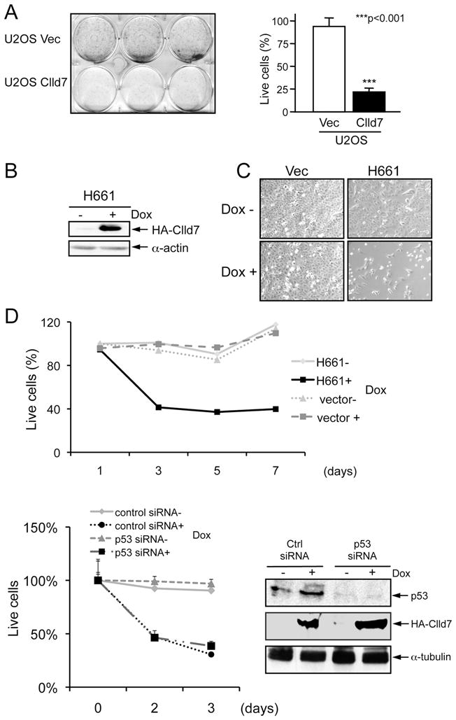
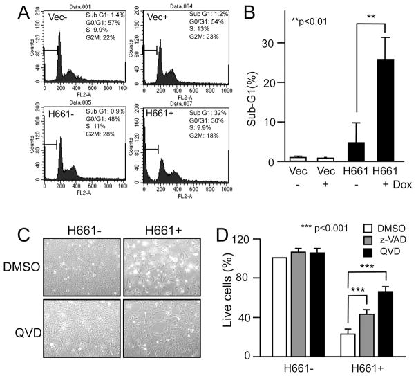
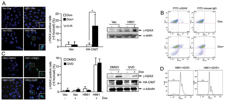
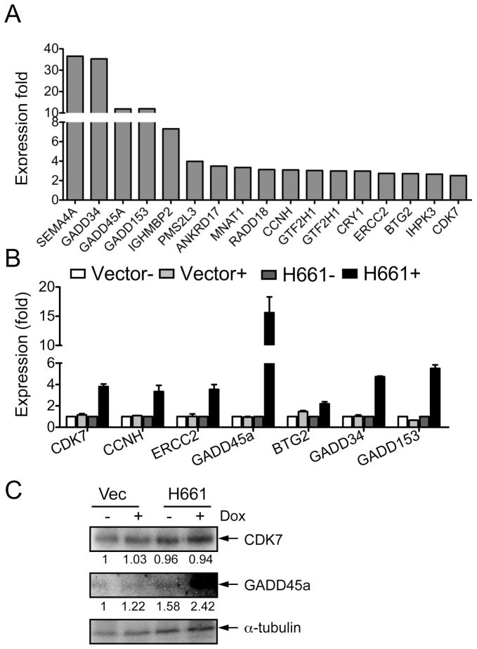
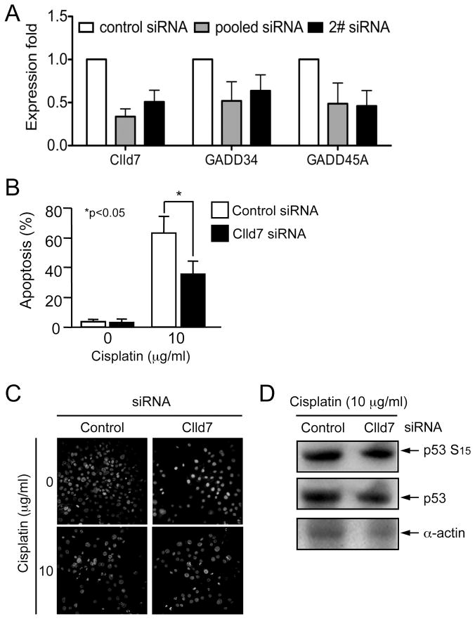
Chronic lymphocytic leukemia deletion gene 7 (Clld7) is a candidate tumor suppressor on chromosome 13q14. Clld7 encodes an evolutionarily conserved protein that contains an RCC1 domain plus broad complex, tramtrack, bric-a-brac (BTB), and POZ domains. In this study, we investigated the biological functions of Clld7 protein in inducible osteosarcoma cell lines. Clld7 induction inhibited cell growth, decreased cell viability, and increased γ-H2AX staining under conditions of caspase inhibition, indicating activation of the DNA damage/repair pathway. Real-time PCR analysis in tumor cells and normal human epithelial cells revealed Clld7 target genes that regulate DNA repair responses. Furthermore, depletion of Clld7 in normal human epithelial cells conferred resistance to apoptosis triggered by DNA damage. Taken together, the biological actions of Clld7 are consistent with those of a tumor suppressor.


Figures

Figure 1
Figure 1
Real-time PCR analysis of Clld7 expression in tumor lines. Clld7 expression in each tumor cell line (black bars) is indicated by its ratio to that of the corresponding normal tissues (white bars). GAPDH was used as the reference.
Figure 2
Figure 2
Suppression of cell proliferation by Clld7 in U2OS cells. A. Overexpression of Clld7 decreases colony formation in U2OS cells. After 10 days of G418 selection, live cells were stained with sulforhodamine B (SRB). In the left panel, wells in the upper row are cells transfected with vector; wells in the lower row are cells transfected with Clld7. Right panel is a bar graph depicting the percentage of live cells in the experiment shown in the left panel. Results represent averages and standard deviations from triplicate wells. B. Inducible expression of Clld7 in H661, a clonal U2OS tet on line expressing C-terminal HA epitope tagged Clld7, as assessed by Western blotting. α-actin was used as a loading control. C. Decreased H661 cell density after 3 days of Clld7 expression by doxycycline (Dox) treatment (2 μg/ml). Treatments with and without Dox are indicated by + and −. D. Cell viability of H661 and U2OS tet on-vector cells as assessed by AlamarBlue staining. Upper panel: H661 cells were treated with Dox for the indicated times. Lower Panel: H661 cells were transfected with p53 specific siRNA or a non-targeting control siRNA followed by Dox treatment for the indicated times. Efficiency of p53 depletion and Clld7 expression were confirmed by Western blot (right panel). α-tubulin was used as loading control.
Figure 3
Figure 3
Clld7 overexpression decreases cell viability through induction of apoptosis. A. Cell cycle profile determined by FACS in H661 cells. Representative data from three independent experiments are shown. Percentage of sub-G1 phase is graphed in B. C. Pan-caspase inhibitor QVD (20 μM) abrogates Dox mediated Clld7 expression in H661 cells. D. Cell viability in C is determined by AlamarBlue staining. Caspase inhibitors z-VAD (50 μM) and QVD (20 μM) were added at 12 hours after Dox treatment for 48 hours. Cell viability in H661 cells without Dox and caspase inhibitor treatment is defined as 100%.
Figure 4
Figure 4
Clld7 induced γ-H2AX foci in U2OS cells. A. Left panel: representative immunofluorescence images showing accumulation of γ-H2AX foci (green) upon inducible expression of Clld7 (H661+). Empty vector transfected cells are shown as a control (Vec− and Vec+). Inserted pictures are zoomed-in images. Middle panel: quantification of γ-H2AX positive cells (>5 foci/cell) in H661+/− and Vec+/−. ~300 cells were counted for each group. Averages and standard deviations are from three independent experiments. Right panel: Western blot analysis of γ-H2AX in H661+/− and Vec+/− cells. B. Live cells stained with γ-H2AX are quantified in H661 upon Dox mediated Clld7 induction by FACS. H661 cells +/− Dox treatment were stained with FITC labeled γ-H2AX antibodies or FITC labeled mouse IgG as a control. Cells were co-stained with Propidium iodide to determine DNA content. C. Immunofluorescence images of γ-H2AX foci (green) (left panel) and quantification of γ-H2AX staining (middle panel) in H661 cells with or without Dox and caspase inhibitor QVD (20 μM) treatment. Approximately 300 cells were counted for each group. Averages and standard deviation are from three independent experiments. Right panel, Western blot analysis of γ-H2AX and inducible expression of HA-Clld7 in H661 cells with and without QVD treatment. D. Flow cytometry analysis of cells used in C after cells were stained with Propidium iodide.
Figure 5
Figure 5
Overexpression of Clld7 deregulates expression of genes in the DNA damage/repair pathway. A. PCR-array analysis documents 17 genes with increased expression upon inducible expression of Clld7 in H661 cells. B. Real-time PCR documents increased expression of 7 genes in H661+ cells as compared to H661- cells. Vector transfected cells were used as control. C. Increased GADD45A protein expression upon Dox-induced expression of Clld7 in H661 cells as assessed by Western blot. α–tubulin was used as a loading control.
Figure 6
Figure 6
Depletion of Clld7 in primary human keratinocytes increases resistance to cisplatin-induced apoptosis. A. Decreased expression of GADD45A and GADD34 after Clld7 depletion. Human primary foreskin keratinocytes (HFKs) were transfected with a Clld7 specific siRNA pool, an individual Clld7 specific siRNA, or control siRNA. Five days after transfection, RNA was extracted for real-time PCR detection. B. HFKs transfected with a Clld7 specific siRNA pool or a control siRNA were treated with the chemotherapy agent cisplatin (10μg/ml) for 23 hours and stained with Hochest33258. A minimum 1,000 cells were counted blindly in each group from three independent populations of HFKs. C. Representative images of HFKs from B. D. HFKs are transfected with non-targeting control and Clld7 siRNA followed by cisplatin treatment (10 μg/ml) for 23 hours. Cell lysates were analyzed by western blot for the expression of Serine 15-phosphorylated p53 and total p53. α–actin was used as a loading control.

References

    1. Ripolles L, Ortega M, Ortuno F, et al. Genetic abnormalities and clinical outcome in chronic lymphocytic leukemia. Cancer Genet Cytogenet. 2006;171:57–64. - PubMed
    1. Cotter FE, Auer RL. Genetic alteration associated with chronic lymphocytic leukemia. Cytogenet Genome Res. 2007;118:310–9. - PubMed
    1. Wada M, Okamura T, Okada M, et al. Delineation of the frequently deleted region on chromosome arm 13q in B-cell non-Hodgkin’s lymphoma. Int J Hematol. 2000;71:159–66. - PubMed
    1. Wada M, Okamura T, Okada M, et al. Frequent chromosome arm 13q deletion in aggressive non-Hodgkin’s lymphoma. Leukemia. 1999;13:792–8. - PubMed
    1. Liu Y, Hermanson M, Grander D, et al. 13q deletions in lymphoid malignancies. Blood. 1995;86:1911–5. - PubMed

Publication types

MeSH terms

Substances