. 2010 Dec;84(24):13004-18.
doi: 10.1128/JVI.01255-10. Epub 2010 Oct 6.

Metagenomic analysis of the viromes of three North American bat species: viral diversity among different bat species that share a common habitat

Affiliations

Metagenomic analysis of the viromes of three North American bat species: viral diversity among different bat species that share a common habitat

Eric F Donaldson et al. J Virol. 2010 Dec.

Abstract

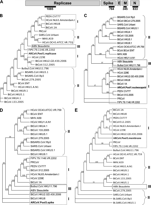
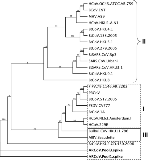
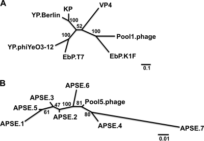
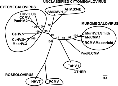
Effective prediction of future viral zoonoses requires an in-depth understanding of the heterologous viral population in key animal species that will likely serve as reservoir hosts or intermediates during the next viral epidemic. The importance of bats as natural hosts for several important viral zoonoses, including Ebola, Marburg, Nipah, Hendra, and rabies viruses and severe acute respiratory syndrome-coronavirus (SARS-CoV), has been established; however, the large viral population diversity (virome) of bats has been partially determined for only a few of the ∼1,200 bat species. To assess the virome of North American bats, we collected fecal, oral, urine, and tissue samples from individual bats captured at an abandoned railroad tunnel in Maryland that is cohabitated by 7 to 10 different bat species. Here, we present preliminary characterization of the virome of three common North American bat species, including big brown bats (Eptesicus fuscus), tricolored bats (Perimyotis subflavus), and little brown myotis (Myotis lucifugus). In samples derived from these bats, we identified viral sequences that were similar to at least three novel group 1 CoVs, large numbers of insect and plant virus sequences, and nearly full-length genomic sequences of two novel bacteriophages. These observations suggest that bats encounter and disseminate a large assortment of viruses capable of infecting many different animals, insects, and plants in nature.


Figures

FIG. 1.
FIG. 1.
Distribution of sequence read matches by pool. The 576,274 sequence reads were compared to the viral RefSeq database using tblastx with an E-value limit of 10e−04 to determine which reads were associated with which viruses. Each pool described in Table 2 is represented as a pie chart, with the percentage of viruses for each category colored according to the key.
FIG. 2.
FIG. 2.
Novel group 1 CoV phylogeny. At least three novel group 1 CoVs were identified in this study as CoV sequences were found in three pools (pools 1 to 3). (A) The CoV genome is comprised of five genes found in all CoVs, including the replicase that makes up the first two-thirds of the genome, and four structural genes including the spike (S), the envelope (E), the membrane (M), and the nucleocapsid (N) genes. Black bars represent the sequences identified and compared in this study. Maximum-likelihood analyses were conducted using the CoV contig sequences identified in this study in comparison to known CoV reference genomes. (B) Pool 1 contained a contig of 1,148 nt that was compared to other CoV reference genomes. An alignment of the most conserved 1,085 nt suggests that this contig is most closely related to group 1 HCoV-229E. (C) A second 833-nt contig in pool 1 matched most closely to the nucleocapsid gene of BtCoV-1A. (D) A 764-nt contig in pool 3 matched most closely to the spike gene of BtCoV-HKU8, a group 1 CoV. (E) A 450-nt contig in pool 3 was most closely related to the membrane gene of BtCoV-HKU2. Values at branch points represent bootstrap values based upon 100 replicates. Branches with values of <70 should be interpreted with caution. Trees are shown as proportional cladograms. Classic CoV group numbers are shown as roman numerals. Bat viruses are indicated with the prefix Bt. HCoV indicates human virus. MHV, murine hepatitis virus; FIPV, feline infectious peritonitis virus; AIBV, avian infectious bronchitis virus; FIPV, feline infectious peritonitis virus; BCoV, bovine coronovirus; PRCoV, porcine respiratory coronovirus.
FIG. 3.
FIG. 3.
Variation between CoV spike genes found in different pools. A sequence read of 412 nt in length from pool 1, Pool 1.287740, matched to the larger spike gene fragment assembled from pool 3, indicating that two related CoVs were found in pools 1 and 3. This 412-nt region shared 96.4% pairwise similarity at the nucleotide level and 98.2% pairwise identity at the amino acid level between the pool 1 and pool 3 variants, and both were most closely related to BtCoV-HKU2. The tree was generated via maximum-likelihood analysis, with 100 bootstrap replicates, and is shown as a proportional cladogram.
FIG. 4.
FIG. 4.
Amplification of the replicase fragment of ARCoV from pool 1. (A) A total of 45 sequence reads derived from 454 sequencing of the pool 1 sample assembled to the BtCoV-HKU2 genome, and these were used to guide PCR amplification of a 2,540-nt fragment of ORF1ab of ARCoV. Green, 454 reads; blue, amplicons derived by PCR and Sanger sequencing; gray, replicase gene; pink, structural and accessory genes. (B) Phylogenetic analysis of the 2,540-nt fragment of the replicase shows that the ARCoV from pool 1 is most closely related to the group 1 CoVs, with its nearest neighbor being BtCoV-HKU2. (C) Phylogenetic analysis of the 833 amino acids suggests the ARCoV replicase sequence is most closely related to HCoV-229E. Trees are shown as proportional cladograms. Values at branch points represent bootstrap values based upon 100 replicates. Branches with values of <70 should be interpreted with caution. Classic CoV group numbers are shown as roman numerals.
FIG. 5.
FIG. 5.
Novel insect viruses of the Iflaviridae family. Six unique 1,449-nt fragments from contigs assembled using sequence from four different pools (pools 1, 2 [n = 3], 4, and 5) were aligned to known insect viruses of the Iflaviridae and Dicistroviridae families and compared by maximum-likelihood analysis. Phylogenetic analysis demonstrated that these novel sequences clustered with other viruses of the Iflaviridae family, with all being most closely related to SBV and DWV, both of which are viruses of honeybees. Abbreviations are as follows: InV, insect virus; KBV, Kashmir bee virus; ALPV, aphid lethal paralysis virus; BQCV, black queen colony virus; CPV, cricket paralysis virus. Values at branch points represent bootstrap values based upon 100 replicates. Branches with values of <70 should be interpreted with caution. Scale bar represents 1 substitution per nucleotide position.
FIG. 6.
FIG. 6.
Novel plant viruses of the Tymoviridae family. Two nearly identical plant virus sequences were identified in pool 1 and pool 3, and a 1,902-nt segment of each was most closely related to viruses of the Tymoviridae family. These viruses appear to form a new genus in the family as a sister clade to the Maculavirus genus. Abbreviations are as follows: PlV, plant virus; GFkV, grapevine fleck virus; CSDV, citrus sudden death-associated virus; OBDV, oat blue dwarf virus; KYMV, Kennedia yellow mosaic virus; OMV, okra mosaic virus; TYMV, turnip yellow mosaic virus; EMV, eggplant mosaic virus; PhyMV, Physalis mosaic virus. Numbers at branch points represent bootstrap values based upon 100 iterations. Scale bar represents 0.01 substitutions per nucleotide site.
FIG. 7.
FIG. 7.
Two novel bacteriophage sequences identified in different pools. Several pools contained sequences that matched most closely to different bacteriophages. (A) In pool 1, a phage most closely related to enterobacteria phage (EbP) K1F was identified, and a phylogenetic tree was generated using maximum likelihood to compare the DNA polymerase gene of the novel pool 1 phage to that of other members of the T7-like genus. The tree demonstrates that the novel phage is most closely related to EbP-K1F. KP, Kluyvera phage Kvp1; VP, vibriophage VP4; YP, Yersinia phage. Values at branch points represent bootstrap values based upon 100 replicates. Branches with values of <70 should be interpreted with caution. Scale bar represents 0.1 substitutions per nucleotide site. (B) In pool 5, the predominant bacteriophage sequence was most closely related to Acyrthosiphon pisum secondary endosymbiont 1 (APSE-1) bacteriophage. The full-length terminase gene was used to compare the novel phage sequence to seven previously described APSE phage sequences. The phylogenetic analysis shows that the pool 5 APSE-like sequence is a novel bacteriophage most closely related to APSE-4 and APSE-6. Values at branch points represent bootstrap values based upon 100 replicates. Branches with values of <70 should be interpreted with caution. Scale bar represents 0.01 substitutions per nucleotide site.
FIG. 8.
FIG. 8.
Phylogeny of a novel herpesvirus found in pool 6. Several contigs from pool 6 matched most closely to viruses in a variety of genera of the Betaherpesvirinae subfamily. A 267-amino acid fragment found in pool 6 was aligned and compared to sequences of several reference viruses of the Betaherpesvirinae. Phylogenetic analyses indicated that this amino acid sequence may be from a novel bat cytomegalovirus that forms a new genera between Roseolovirus and Muromegalovirus. HHV, human herpesvirus; CCMV, chimpanzee cytomegalovirus; PanHV, panine herpesvirus; CeHV, cercopithecine herpesvirus; MacHV, macacine herpesvirus; PCMV, porcine cytomegalovirus; TuHV, tupaiid herpesvirus; RCMV, rat cytomegalovirus; MoCMV, mouse cytomegalovirus; MurHV, murid herpesvirus; AHV, atoline herpesvirus; SQCMV, squirrel monkey cytomegalovirus. Values at branch points represent bootstrap values based upon 100 replicates. Branches with values of <70 should be interpreted with caution. Scale bar represents 0.1 substitutions per nucleotide site.

References

    1. Allander, T., S. U. Emerson, R. E. Engle, R. H. Purcell, and J. Bukh. 2001. A virus discovery method incorporating DNase treatment and its application to the identification of two bovine parvovirus species. Proc. Natl. Acad. Sci. U. S. A. 98:11609-11614. - PMC - PubMed
    1. Allander, T., T. Jartti, S. Gupta, H. G. Niesters, P. Lehtinen, R. Osterback, T. Vuorinen, M. Waris, A. Bjerkner, A. Tiveljung-Lindell, B. G. van den Hoogen, T. Hyypia, and O. Ruuskanen. 2007. Human bocavirus and acute wheezing in children. Clin. Infect. Dis. 44:904-910. - PMC - PubMed
    1. Allander, T., M. T. Tammi, M. Eriksson, A. Bjerkner, A. Tiveljung-Lindell, and B. Andersson. 2005. Cloning of a human parvovirus by molecular screening of respiratory tract samples. Proc. Natl. Acad. Sci. U. S. A. 102:12891-12896. - PMC - PubMed
    1. Anderson, N. G., J. L. Gerin, and N. L. Anderson. 2003. Global screening for human viral pathogens. Emerg. Infect. Dis. 9:768-774. - PMC - PubMed
    1. Arias, C. F., M. Escalera-Zamudio, L. Soto-Del Rio Mde, A. G. Cobian-Guemes, P. Isa, and S. Lopez. 2009. Molecular anatomy of 2009 influenza virus A (H1N1). Arch. Med. Res. 40:643-654. - PubMed

Publication types

MeSH terms

LinkOut - more resources