

Elevated circulating IGF-I promotes mammary gland development and proliferation
- PMID: 20926579
- PMCID: PMC2999497
- DOI: 10.1210/en.2010-0792
Elevated circulating IGF-I promotes mammary gland development and proliferation
Abstract
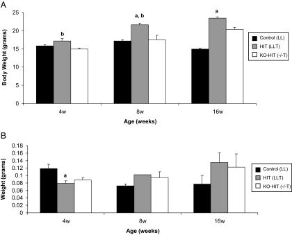
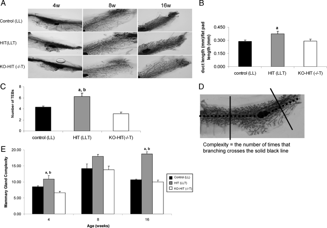
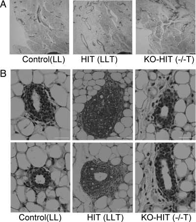
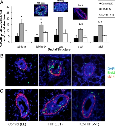
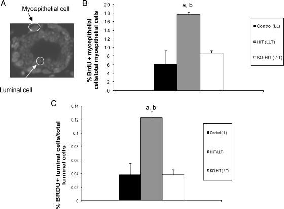
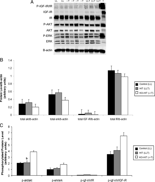
Animal studies have shown that IGF-I is essential for mammary gland development. Previous studies have suggested that local IGF-I rather than circulating IGF-I is the major mediator of mammary gland development. In the present study we used the hepatic IGF-I transgenic (HIT) and IGF-I knockout/HIT (KO-HIT) mouse models to examine the effects of enhanced circulating IGF-I on mammary development in the presence and absence of local IGF-I. HIT mice express the rat IGF-I transgene under the transthyretin promoter in the liver and have elevated circulating IGF-I and normal tissue IGF-I levels. The KO-HIT mice have no tissue IGF-I and increased circulating IGF-I. Analysis of mammary gland development reveals a greater degree of complexity in HIT mice as compared to control and KO-HIT mice, which demonstrate similar degrees of mammary gland complexity. Immunohistochemical evaluation of glands of HIT mice also suggests an enhanced degree of proliferation of the mammary gland, whereas KO-HIT mice exhibit mammary gland proliferation similar to control mice. In addition, HIT mice have a higher percentage of proliferating myoepithelial and luminal cells than control mice, whereas KO-HIT mice have an equivalent percentage of proliferating myoepithelial and luminal cells as control mice. Thus, our findings show that elevated circulating IGF-I levels are sufficient to promote normal pubertal mammary epithelial development. However, HIT mice demonstrate more pronounced mammary gland development when compared to control and KO-HIT mice. This suggests that both local and endocrine IGF-I play roles in mammary gland development and that elevated circulating IGF-I accelerates mammary epithelial proliferation.
Figures
Similar articles
-
A moderate elevation of circulating levels of IGF-I does not alter ErbB2 induced mammary tumorigenesis.BMC Cancer. 2011 Aug 25;11:377. doi: 10.1186/1471-2407-11-377. BMC Cancer. 2011. PMID: 21867536 Free PMC article.
-
SirT1 modulates the estrogen-insulin-like growth factor-1 signaling for postnatal development of mammary gland in mice.Breast Cancer Res. 2007;9(1):R1. doi: 10.1186/bcr1632. Breast Cancer Res. 2007. PMID: 17201918 Free PMC article.
-
Developmental stage determines estrogen receptor alpha expression and non-genomic mechanisms that control IGF-1 signaling and mammary proliferation in mice.J Clin Invest. 2012 Jan;122(1):192-204. doi: 10.1172/JCI42204. Epub 2011 Dec 19. J Clin Invest. 2012. PMID: 22182837 Free PMC article.
-
Genetic manipulation of the IGF-I axis to regulate mammary gland development and function.J Dairy Sci. 2002 Feb;85(2):365-77. doi: 10.3168/jds.S0022-0302(02)74083-6. J Dairy Sci. 2002. PMID: 11913696 Review.
-
The insulin-like growth factors (IGFs) and IGF binding proteins in postnatal development of murine mammary glands.J Mammary Gland Biol Neoplasia. 2000 Jan;5(1):31-42. doi: 10.1023/a:1009511131541. J Mammary Gland Biol Neoplasia. 2000. PMID: 10791766 Review.
Cited by
-
Insulin-like growth factor-I receptor (IGF-IR) translocates to nucleus and autoregulates IGF-IR gene expression in breast cancer cells.J Biol Chem. 2012 Jan 20;287(4):2766-76. doi: 10.1074/jbc.M111.281782. Epub 2011 Nov 29. J Biol Chem. 2012. PMID: 22128190 Free PMC article.
-
The Insulin-like Growth Factor Signaling Pathway in Breast Cancer: An Elusive Therapeutic Target.Life (Basel). 2022 Nov 29;12(12):1992. doi: 10.3390/life12121992. Life (Basel). 2022. PMID: 36556357 Free PMC article. Review.
-
A moderate elevation of circulating levels of IGF-I does not alter ErbB2 induced mammary tumorigenesis.BMC Cancer. 2011 Aug 25;11:377. doi: 10.1186/1471-2407-11-377. BMC Cancer. 2011. PMID: 21867536 Free PMC article.
-
Type I insulin-like growth factor receptor signaling in hematological malignancies.Oncotarget. 2017 Jan 3;8(1):1814-1844. doi: 10.18632/oncotarget.12123. Oncotarget. 2017. PMID: 27661006 Free PMC article. Review.
-
Elevated GH/IGF-I promotes mammary tumors in high-fat, but not low-fat, fed mice.Carcinogenesis. 2014 Nov;35(11):2467-73. doi: 10.1093/carcin/bgu161. Epub 2014 Aug 1. Carcinogenesis. 2014. PMID: 25085903 Free PMC article.
References
-
- Richert MM, Schwertfeger KL, Ryder JW, Anderson SM 2000 An atlas of mouse mammary gland development. J Mammary Gland Biol Neoplasia 5:227–241 - PubMed
-
- Kleinberg DL 1998 Role of IGF-I in normal mammary development. Breast Cancer Res Treat 47:201–208 - PubMed
-
- Wood TL, Richert MM, Stull MA, Allar MA 2000 The insulin-like growth factors (IGFs) and IGF binding proteins in postnatal development of murine mammary glands. J Mammary Gland Biol Neoplasia 5:31–42 - PubMed
-
- LeRoith D, Neuenschwander S, Wood TL, Hennighausen L 1995 Insulin-like growth factor-I and insulin-like growth factor binding protein-3 inhibit involution of the mammary gland following lactation: studies in transgenic mice. Prog Growth Factor Res 6:433–436 - PubMed
Publication types
MeSH terms
Substances
Grants and funding
LinkOut - more resources
Full Text Sources
Molecular Biology Databases
Research Materials
