. 2010 Dec;104(6):3568-75.
doi: 10.1152/jn.00415.2010. Epub 2010 Oct 6.

Differing neurophysiologic mechanosensory input from glabrous and hairy skin in juvenile rats

Affiliations

Differing neurophysiologic mechanosensory input from glabrous and hairy skin in juvenile rats

M Danilo Boada et al. J Neurophysiol. 2010 Dec.

Abstract

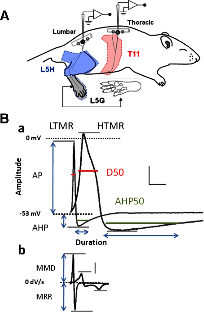
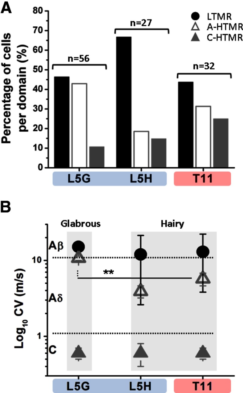
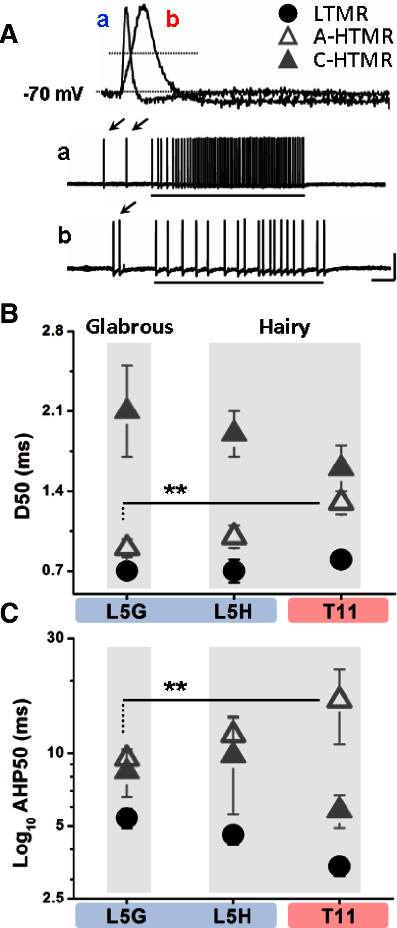
Sensory afferents in skin encode and convey thermal and mechanical conditions, including those that threaten tissue damage. A small proportion of skin, the glabrous skin of the distal extremities, is specialized to explore the environment in fine detail. Aside from increased innervation density, little is known regarding properties of mechanosensory afferents to glabrous skin in younger animals that explain the exquisite precision and high contrast in rapidly sampling physical structures, including those that threaten injury. To assess this, we obtained intact neuronal intracellular recordings in vivo from 115 mechanosensitive afferent neurons from lumbar and thoracic dorsal root ganglia in juvenile rats. Two characteristics were unique to glabrous skin: a threefold higher proportion of fast-conducting to slow-conducting afferents that were high-threshold mechanosensitive nociceptors compared with hairy skin and a twofold faster conduction velocity of fast-conducting nociceptors compared with hairy skin. Additionally differences were found in mechanical thresholds between glabrous skin and hairy skin for each fiber type. These differences reflect and help explain the rapid response of skin specialized to explore the physical environment. Additionally, these results highlight potential limitations of using passive electrical properties and conduction velocity alone to characterize primary afferents without knowledge of the skin type they innervated.


Figures

Fig. 1.
Fig. 1.
A: schematic diagram of the in vivo rat 5th lumbar/11th thoracic (L5/T11) preparation (lateral view). Colored areas delineate approximate dermatome boundaries where skin sensory neuron receptive fields (RFs) were located (L5G: glabrous skin [paw plantar surface]; L5H: hairy skin [Leg]). B: active properties measured in dorsal root ganglia (DRG) neurons. Typical waveforms (tactile [LTMR] and nociceptive [HTMR]) of action potential (AP) (a) and dV/dt (b). Scale bars: 20 mV, 100 V/s, 2 ms. LTMR, low-threshold mechanoreceptor; HTMR, high-threshold mechanoreceptor; AHTMR, Aδ nociceptor HTMR; CHTMR, C-nociceptor HTMR.
Fig. 2.
Fig. 2.
A: population distribution of main cellular subtypes recorded at different ganglia. The percentage of cells of each subtype categorized by dermatome (L5, 5th lumbar; T11, 11th thoracic) and skin type (G, glabrous skin; H, hairy skin). n = total number of cells analyzed in each skin type domain. The ratio of fast-conducting (AHTMR) to slow-conducting high-threshold (CHTMR) mechanoreceptors was threefold greater in the glabrous skin compared with the hairy skin (P < 0.05). Neuron types: LTMR, ▵ AHTMR, ▴ CHTMR. B: relationship between cellular subtype and log10 conduction velocity (CV) at different thoracic and lumbar ganglia (T11 or L5) and for different skin types, hairy or glabrous (H or G). There is a significant difference between CV of AHTMR afferents demonstrated with the line (**P < 0.05). The higher CV of the AHTMR in L5G is clear, making CV alone more difficult to delineate AHTMR from LTMR in glabrous skin or AHTMR in glabrous skin from LTMR in hairy skin in either dermatome. A clear difference in CV distribution of AHTMR and LTMR relative to CHTMR in glabrous skin can be appreciated. This difference from CHTMR is smaller in L5H and T11. Neuron types: LTMR, ▵ AHTMR, ▴ CHTMR.
Fig. 3.
Fig. 3.
A: representative AP shape of typical fast nociceptors from glabrous skin (at L5) (a, blue) and hairy skin (at T11) (b, red). Responses to suprathreshold stimulation (15 mN) are also shown. Both cells have similar mechanical threshold (MT) and CV (±7 m/s). Arrows point to the initial contact of the VF with the RF of the cells before applying full force. The clear differences in somatic electrical properties of the fast nociceptors from glabrous and hairy skin are readily visualized. Scale bars: AP traces 20 mV, 1.3 ms. Response traces 30 mV, 0.3 s. B: relationship between AP duration (at 50% of amplitude [D50]) and cellular subtype in both ganglia. A significant difference was found between AHTMR in glabrous skin in L5 and T11 (**P < 0.05), whereas there was overlap between AHTMR and CHTMR at T11. C: relation between cellular subtype and log10 AHP50 (log of afterhyperpolarization duration at 50% of amplitude) at different ganglia and skin type. A significant difference was found between the AHTMR from L5 and T11 (**P < 0.05). However, there was significant overlap between AHTMR and CHTMR in the neurons from the L5 ganglia. Neuron types: LTMR, ▵ AHTMR, ▴ CHTMR. AP, action potential; CV, conduction velocity; VF, von Frey filament; RF, receptive field; mV, millivolts; mN, millinewtons; ms, milliseconds.

References

    1. Albers KM, Woodbury CJ, Ritter AM, Davis BM, Koerber HR. Glial cell-line-derived neurotrophic factor expression in skin alters the mechanical sensitivity of cutaneous nociceptors. J Neurosci 26: 2981–2990, 2006 - PMC - PubMed
    1. Bennett GJ, Chung JM, Honore M, Seltzer Z. Models of neuropathic pain in the rat. Curr Protoc Neurosci May: Unit 9.14, 2003 - PubMed
    1. Bessou P, Perl ER. Response of cutaneous sensory units with unmyelinated fibers to noxious stimuli. J Neurophysiol 32: 1025–1043, 1969 - PubMed
    1. Birznieks I, Jenmalm P, Goodwin AW, Johansson RS. Encoding of direction of fingertip forces by human tactile afferents. J Neurosci 21: 8222–8237, 2001 - PMC - PubMed
    1. Blake DT, Byl NN, Merzenich MM. Representation of the hand in the cerebral cortex. Behav Brain Res 135: 179–184, 2002 - PubMed

Publication types

LinkOut - more resources