Distinguishing Ménétrier's disease from its mimics
- PMID: 20926644
- PMCID: PMC3020399
- DOI: 10.1136/gut.2010.220061
Distinguishing Ménétrier's disease from its mimics
Abstract
Objective: Ménétrier's disease (MD) is a rare hypertrophic gastropathy characterised by giant rugal folds, hypochlorhydria, protein loss and a classic constellation of symptoms (nausea, vomiting, abdominal pain and peripheral oedema). It is considered a clinical diagnosis that may at times be difficult to establish. Firm diagnostic criteria for MD are proposed by delineating the clinicopathological features that best differentiate MD from its mimics.
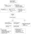
Method: 48 patients referred to Vanderbilt University Medical Center for consideration of enrolment in a clinical trial of treatment of patients with MD with cetuximab were evaluated for a definitive diagnosis by assessing the clinical presentation, pertinent laboratory values and histopathological features.
Results: MD was confirmed in 25 of the 48 patients (52%). The remaining 23 patients were considered to be mimics of MD, the most common diagnoses being gastric polyps or polyposis syndromes (13/23, 57%). Gastric slides were available from 40 of the 48 patients for detailed histological analysis (22/25 MD and 18/23 non-MD). Foveolar hyperplasia, glandular tortuosity and dilation, and a marked reduction in parietal cell number were present in all 22 cases of MD. Lamina propria smooth muscle hyperplasia and oedema characterised most cases (18/22 and 19/22, respectively). More than half had prominent eosinophils (11/22) and/or plasma cells (12/22) in the lamina propria. The clinical presentation of patients with MD was characterised by significantly younger age of onset, male predominance and increased vomiting compared with non-MD patients, and a lower prevalence of anaemia compared with MD patients with polyps. There was a trend towards increased frequency of peripheral oedema in patients with MD compared with non-MD patients.
Conclusions: MD is most accurately diagnosed by clinicohistopathological analysis including oesophagogastroduodenoscopy with gastric pH, appropriate laboratory tests (complete blood count, serum albumin, serum gastrin, Helicobacter pylori and cytomegalovirus serology) and full-thickness mucosal biopsy of the involved gastric mucosa.
Conflict of interest statement
Competing Interest
None to declare.
Figures


References
-
- Ménétrier P. Des polyadenomes gastriques et leur rapport avec le cancer de l’estomac. Arch Physiol Norm Pathol. 1888;1:236–62.
-
- Scharschmidt B. The natural history of hypertrophic gastrophy (Ménétrier’s disease). Report of a case with 16 year follow-up and review of 120 cases from the literature. Am J Med. 1977;63:644–62. - PubMed
-
- Drut RM, Gomez MA, Lojo MM, et al. Cytomegalovirus-associated Menetrier’s disease in adults. Demonstration by polymerase chain reaction (PCR) Medicina (B Aires) 1995;55:659–64. - PubMed
-
- Eisenstat DD, Griffiths AM, Cutz E, et al. Acute cytomegalovirus infection in a child with Ménétrier’s disease. Gastroenterology. 1995;109:592–95. - PubMed
Publication types
MeSH terms
Grants and funding
LinkOut - more resources
Full Text Sources
