. 2010 Oct 6;30(40):13441-53.
doi: 10.1523/JNEUROSCI.0907-10.2010.

Quantitative analysis of synaptic vesicle Rabs uncovers distinct yet overlapping roles for Rab3a and Rab27b in Ca2+-triggered exocytosis

Affiliations

Quantitative analysis of synaptic vesicle Rabs uncovers distinct yet overlapping roles for Rab3a and Rab27b in Ca2+-triggered exocytosis

Nathan J Pavlos et al. J Neurosci. .

Abstract

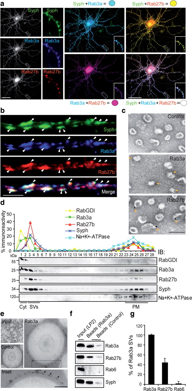
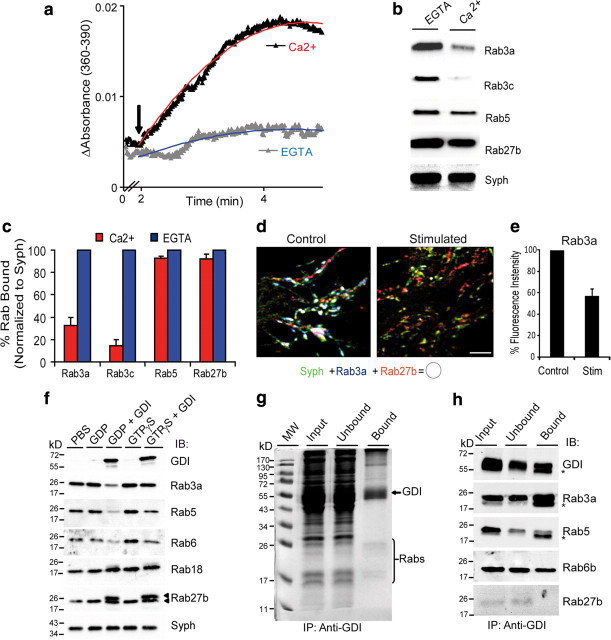
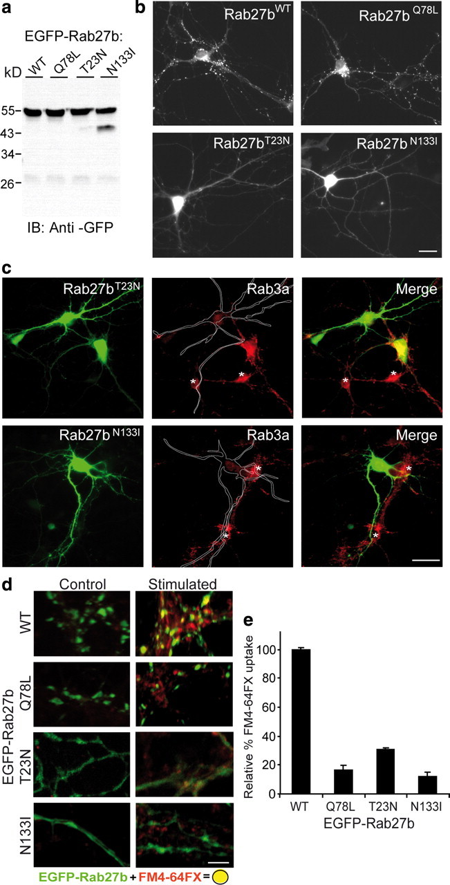
Rab GTPases are molecular switches that orchestrate protein complexes before membrane fusion reactions. In synapses, Rab3 and Rab5 proteins have been implicated in the exo-endocytic cycling of synaptic vesicles (SVs), but an involvement of additional Rabs cannot be excluded. Here, combining high-resolution mass spectrometry and chemical labeling (iTRAQ) together with quantitative immunoblotting and fluorescence microscopy, we have determined the exocytotic (Rab3a, Rab3b, Rab3c, and Rab27b) and endocytic (Rab4b, Rab5a/b, Rab10, Rab11b, and Rab14) Rab machinery of SVs. Analysis of two closely related proteins, Rab3a and Rab27b, revealed colocalization in synaptic nerve terminals, where they reside on distinct but overlapping SV pools. Moreover, whereas Rab3a readily dissociates from SVs during Ca(2+)-triggered exocytosis, and is susceptible to membrane extraction by Rab-GDI, Rab27b persists on SV membranes upon stimulation and is resistant to GDI-coupled Rab retrieval. Finally, we demonstrate that selective modulation of the GTP/GDP switch mechanism of Rab27b impairs SV recycling, suggesting that Rab27b, probably in concert with Rab3s, is involved in SV exocytosis.


Figures

Figure 1.
Figure 1.
Identification and quantitation of Rab GTPases in rat brain homogenates by iTRAQ labeling and tandem MS. a, EM characterization of negatively stained samples of starting brain homogenate, synaptic cytosol, crude SVs, and highly purified SVs. Scale bar, 0.5 μm. b, A schematic of the rat SV purification protocol. Rat brains were homogenized and then centrifuged in two low-speed steps to generate a crude synaptosomal particulate fraction (P2′) and cytosolic fraction (S3). Following osmotic lysis of the synaptosomes and two low-speed centrifugations, crude SVs were further purified by sucrose density centrifugation and size-exclusion chromatography (SEC) on controlled pore glass beads (CPG). Peak fractions of SVs were pooled and collected by centrifugation to yield a highly purified SV fraction. Fractions were digested with trypsin and the peptides labeled with 114, 115, 116, and 117 iTRAQ reagents, respectively. Labeled peptides were then prefractionated off-line by SCX chromatography and analyzed by reverse-phase LC-MS. MS/MS spectra of iTRAQ-labeled peptides from Rab3a and Rab18 are illustrated. The peak areas of reporter ions 114, 115, 116, and 117 in the MS/MS spectra correspond to the relative abundance of the Rab proteins in the various brain homogenates. c, Quantitative proteomic analysis of Rabs in isolated rat brain homogenates. Data represent the means ± SD of iTRAQ ratios from two biological replicates and are expressed as relative abundance over starting homogenate. Segmented line denotes enrichment threshold.
Figure 2.
Figure 2.
Enrichment profiles of Rab GTPases on purified SVs isolated from rat brain homogenates, determined by quantitative immunoblotting. a, Immunoblot analysis of various membrane and soluble rat brain fractions obtained during the purification of SVs by differential centrifugation and controlled pore glass chromatography. Five micrograms of each fraction were resolved by SDS-PAGE (12.5%), transferred to nitrocellulose membrane, and probed overnight with antibodies directed against the indicated proteins. Note that Rab3 proteins and Rab27b exhibit strong coenrichment with SV markers such as Syph and synaptobrevin (Sybrv) but not with plasma membrane protein NMDAR or with soluble cytosolic proteins Rab-GDI and early endosome antigen (EEA1). Each panel is representative of a minimum of three independent blots. b, Quantitative distribution profiles of Rab proteins assigned to functions in distinct organelles (secretory, endosomal, Golgi). Solid lines represent the means of the enrichments (±SD) over homogenate of three independent blots. Segmented lines correspond to the synaptophysin enrichment profile (red) and threshold (black), respectively.
Figure 3.
Figure 3.
A survey of the subcellular localization of SV-enriched Rabs in rat hippocampal neurons. pEGFP-C2-Rab3a, -Rab4b, -Rab5a, -Rab10, -Rab11b, -Rab14, and -Rab27b were each transiently transfected into primary rat hippocampal neurons (10–12 DIV). Forty-eight hours after transfection, neurons were fixed and immunostained with antibodies against Syph (red) and MAP2 (blue) to visualize synaptic boutons and dendrites, respectively. Areas of overlap appear yellow. Arrows highlight colabeled structures. Scale bar, 20 μm.
Figure 4.
Figure 4.
Quantitative immunofluorescence analysis of SV Rabs in rat hippocampal nerve terminals. a, Representative axons of hippocampal neurons (10–12 DIV) transfected with the indicated EGFP-Rab construct and fixed 48 h after transfection. Neurons were subsequently double labeled with antibodies against Rab3a and Syph to visualize synaptic boutons, and correlative line scans were performed across the entire length of the axons with corresponding histograms illustrated below. Scale bar, 5 μm. b, Cross-correlation analyses of EGFP-Rabs with either Rab3a (black bars) or synaptophysin (gray bars) on individual synaptic boutons (n = 50). Secretory Rabs Rab3a (Rr = 0.956) and Rab27b (Rr = 0.828) showed strong correlation with the respective SV markers, whereas Rab35 (Rr = 0.310) showed no obvious correlation. Data represents the means ± SD.
Figure 5.
Figure 5.
Rab27b occupies distinct but overlapping SV pools with Rab3a. a, Immunofluorescence analysis of Rab27b-, Rab3a-, and synaptophysin-labeled synaptic boutons. Hippocampal neurons (17 DIV) were fixed in 3% PFA and triple labeled with antibodies against Rab27b, Rab3a, and Syph. Insets represent magnifications of boxed regions. Colocalization appears white following the overlay of all three fluorescent channels. Scale bar, 10 μm. b, Confocal micrograph of an ultrathin section of a neuronal axon triple labeled with Rab3a, Rab27b, and Syph antibodies. Fluorescently labeled neurons grown on coverslips were postfixed with 2% glutaraldehyde and embedded in 2,4,6-tris[bis(methoxymethyl)amino]-1,3,5-triazine, and ultrathin sections (∼100 nm) were cut before sections were mounted for analysis by confocal microscopy. Arrows denote colocalized puncta. Note that the colocalization between Rab27b and Rab3a is incomplete in the merged panel. c, Immunogold localization of Rab3a and Rab27b on highly purified SVs. SV samples were fixed and incubated in the presence of either rabbit IgG (control), or antibodies against Rab3a or Rab27b. The samples were then incubated with appropriate secondary antibodies conjugated to 10 nm gold. Gold particles (yellow arrows) are distributed on the surface of select SVs. Scale bar, 200 nm. d, Velocity density gradient centrifugation analyses of subfractionated synaptosomes. Gradient fractions (0.4–1.2 m sucrose) were analyzed from top to bottom by quantitative immunoblotting of membranes for the SV marker Syph, Rab3a, Rab27b, cytosolic (Cyto) GDI, and the PM marker Na+/K+-ATPase. e, Presence of Rab27b on SVs immunoisolated with Rab3a. SVs (LP2) were incubated with Eupergit C1Z beads containing covalently attached monoclonal antibodies to Rab3a (Cl 42.2) or incubated with beads whose functional groups had been inactivated with glycine (control). SV binding was monitored by EM morphometry. Inset denotes magnification of boxed region. Scale bar, 500 nm. The co-occupancy of Rabs on Rab3a-immunoisolated SVs was analyzed by immunoblotting (f), quantified (g), and normalized to the total captured Rab3a SV pool. Means ± SEM from three to five independent measurements are shown.
Figure 6.
Figure 6.
Differential membrane dynamics between Rab27b and Rab3a during Ca2+-triggered exocytosis. a, Ca2+-dependent stimulation of glutamate release by K+ depolarization from isolated rat synaptosomes. Synaptosomes (1 mg) were resuspended in sodium buffer containing 1.3 mm calcium chloride, 5 mm EGTA, or 50 mm KCl and glutamate release was monitored spectrophotometrically following the generation of NADPH associated with the conversion of glutamate dehydrogenase. b, Dissociation of Rab3a/c but not Rab5 or Rab27b during Ca2+-triggered exocytosis. Following stimulation and release, synaptosomes were subfractionated to obtain crude SV factions (total assay duration ∼1 h), which were analyzed by SDS-PAGE and immunoblotting with the indicated antibodies. Syph served as a loading control for equivalent amounts of SVs. c, Densitometric analyses of Rabs bound to SVs following Ca2+-triggered exocytosis. Ca2+ and EGTA immunointensities were normalized to their respective Syph controls and expressed as a percentage of the respective EGTA controls. d, Dispersion of Rab3a but not Rab27b upon K+ depolarization. Hippocampal neurons were incubated with neuronal buffer in either the presence (stimulated) or absence (control) of KCl (50 mm) for 1 min, immediately fixed, triple labeled with the indicated antibodies and processed for ultrathin sectioning of synaptic boutons for confocal microscopy. Representative confocal micrographs are depicted. White color denotes Rab3a/Rab27b/Syph colocalization following overlay of individual channels. e, Quantification of the relative Rab3a immunofluorescence intensity in the absence or presence of stimulation (stim). Data are expressed as a percentage of the no stimulation control. f, Rab27b is resistant to GDI-membrane extraction. Isolated SVs (LP2) were incubated for 15 min in the presence of GDP or GTPγS (500 μm) at 37°C. His-GDI (200 nm) or PBS (control) was then added and incubation was continued for an additional 45 min. The membranes were pelleted and then analyzed by SDS-PAGE and immunoblotting. In addition to Rab3a, GDI efficiently removes Rab5 and Rab6 from SV membranes but not Rab18 or Rab27b. Note that a distinct doublet band (arrows) is observed in the Rab27b samples following GDI incubation in both the presence of either GDP or GTPγS. g, Isolation of Rab-GDI complexes by affinity chromatography. Rab-GDI complexes from concentrated presynaptic cytosol (LS2) were captured by an affinity column coupled with a monoclonal antibody specific for GDI. Complexes were eluted by low pH and samples analyzed by immunoblotting (h) and mass spectrometry (see Table 1). Asterisks in g correspond to the light chain of the monoclonal antibody used for affinity purification. Note the low abundance of Rab27b in the starting input and flow through samples and low capture affinity with GDI as compared to other Rabs examined.
Figure 7.
Figure 7.
Functional characterization of Rab27b GTP/GDP-mutants in hippocampal neurons. a, b, Expression and localization of EGFP-Rab27b fusion proteins in rat hippocampal neurons. Hippocampal neurons (8 DIV) were transfected with the indicated EGFP-Rab27b fusion constructs for 48 h and then either harvested for analysis by SDS-PAGE and immunoblotting with an antibody against GFP (a) or fixed and processed for fluorescence microscopy (b). EGFP-Rab27bWT and -Rab27bQ78L mutants localized primarily to synaptic boutons, whereas EGFP-Rab27bT23N and -Rab27bN133I were diffusely distributed throughout the neuronal somas and radiating neurites. c, Overexpression of GDP-Rab27b mutants reduces membrane-bound Rab3a. Hippocampal neurons strongly expressing EGFP-Rab27bT23N and -Rab27bN133I were immunostained with antibodies against endogenous Rab3a and examined by fluorescence microscopy. Overexpression of each EGFP-Rab27b fusion protein correlated with a reduction in Rab3a immunofluorescence levels as compared to untransfected neighboring cells (asterisks). Segmented line outlines boundaries of transfected neurons. d, Expression of Rab27b mutants impairs SV cycling. Confocal images of axonal processes of cultured hippocampal neurons transfected with EGFP-Rab27b wild-type or mutant fusion proteins. The neurons were processed for FM4-64FX uptake (red) in the absence (control) or presence (stimulated) of electrical stimulation (600 APs, 20 Hz) as detailed in the Materials and Methods. Scale bars, 5 μm. e, Bar graph summarizing FM4-64 uptake following transfection with the indicated vectors. The data were accumulated from separate cultures/transfections and expressed as the normalized means of analyzed fields ± SEM (n = 40).

References

    1. Babbey CM, Ahktar N, Wang E, Chen CC, Grant BD, Dunn KW. Rab10 regulates membrane transport through early endosomes of polarized Madin-Darby canine kidney cells. Mol Biol Cell. 2006;17:3156–3175. - PMC - PubMed
    1. Bethani I, Lang T, Geumann U, Sieber JJ, Jahn R, Rizzoli SO. The specificity of SNARE pairing in biological membranes is mediated by both proof-reading and spatial segregation. EMBO J. 2007;26:3981–3992. - PMC - PubMed
    1. Burstein ES, Brondyk WH, Macara IG. Amino acid residues in the Ras-like GTPase Rab3A that specify sensitivity to factors that regulate the GTP/GDP cycling of Rab3A. J Biol Chem. 1992;267:22715–22718. - PubMed
    1. Castillo PE, Schoch S, Schmitz F, Südhof TC, Malenka RC. RIM1alpha is required for presynaptic long-term potentiation. Nature. 2002;415:327–330. - PubMed
    1. Chavas LM, Torii S, Kamikubo H, Kawasaki M, Ihara K, Kato R, Kataoka M, Izumi T, Wakatsuki S. Structure of the small GTPase Rab27b shows an unexpected swapped dimer. Acta Crystallogr D Biol Crystallogr. 2007;63:769–779. - PubMed

Publication types

MeSH terms

LinkOut - more resources