. 2011 Jan 13;117(2):727-35.
doi: 10.1182/blood-2010-05-285718. Epub 2010 Oct 6.

Inhibition of cell adhesion by anti-P-selectin aptamer: a new potential therapeutic agent for sickle cell disease

Affiliations

Inhibition of cell adhesion by anti-P-selectin aptamer: a new potential therapeutic agent for sickle cell disease

Diana R Gutsaeva et al. Blood. .

Abstract

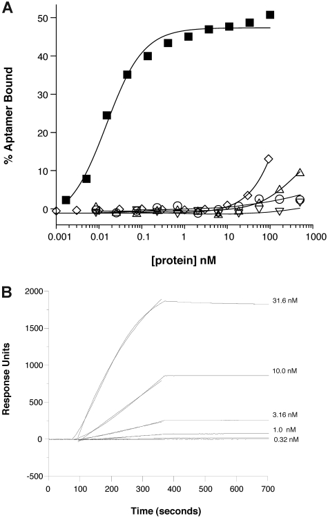
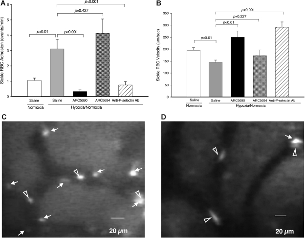
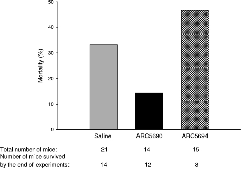
Adhesive interactions between circulating sickle red blood cells (RBCs), leukocytes, and endothelial cells are major pathophysiologic events in sickle cell disease (SCD). To develop new therapeutics that efficiently inhibit adhesive interactions, we generated an anti-P-selectin aptamer and examined its effects on cell adhesion using knockout-transgenic SCD model mice. Aptamers, single-stranded oligonucleotides that bind molecular targets with high affinity and specificity, are emerging as new therapeutics for cardiovascular and hematologic disorders. In vitro studies found that the anti-P-selectin aptamer exhibits high specificity to mouse P-selectin but not other selectins. SCD mice were injected with the anti-P-selectin aptamer, and cell adhesion was observed under hypoxia. The anti-P-selectin aptamer inhibited the adhesion of sickle RBCs and leukocytes to endothelial cells by 90% and 80%, respectively. The anti-P-selectin aptamer also increased microvascular flow velocities and reduced the leukocyte rolling flux. SCD mice treated with the anti-P-selectin aptamer demonstrated a reduced mortality rate associated with the experimental procedures compared with control mice. These results demonstrate that anti-P-selectin aptamer efficiently inhibits the adhesion of both sickle RBCs and leukocytes to endothelial cells in SCD model mice, suggesting a critical role for P-selectin in cell adhesion. Anti-P-selectin aptamer may be useful as a novel therapeutic agent for SCD.


Figures

Figure 1
Figure 1
Primary sequence and proposed secondary structure of anti–mouse P-selectin aptamer ARC5690. ARC5690 is with 40-kDa PEG. idT, inverted 2′-deoxy-thymidine.
Figure 2
Figure 2
Hypoxia/normoxia protocol to study effects of ARC5690 on sickle RBC and leukocyte adhesion. SCD mice were injected with saline, ARC5690 or ARC5694. Two and one-half hours after injection, mice were subjected to 1 hour of hypoxia (FiO2 = 0.12) followed by 1 hour of normoxia in room air (FiO2 = 0.21), and intravital studies were then initiated. During surgery, mice were supplemented with 30% O2. FiO2 indicates fraction of inspired oxygen in a gas mixture.
Figure 3
Figure 3
Anti–P-selectin aptamer binds to murine P-selectin with high specificity. (A) Nitrocellulose filtration assays were performed with 5′-32P-radiolabeled ARC5690 (lacking 5′-PEG) in the presence of increasing concentrations of mouse P-selectin (■), E-selectin (○), L-selectin (▵), or human P-selectin (▿). The aptamer binds to mouse P-selectin with a dissociation constant (KD) of approximately 15pM, and to other proteins with KD > 500nM (the highest protein concentration tested). Radiolabeled scrambled aptamer binds to mouse P-selectin (◇) with KD > 100nM (the highest protein concentration tested). (B) Surface plasmon resonance assay was performed with 5′-biotin–labeled ARC5690 (lacking 5′-PEG) immobilized to a streptavidin-derivatized microfluidics chip. Different concentrations of recombinant murine P-selectin (31.6, 10.0, 3.16, 1.00, and 0.32nM) were injected over the aptamer-coated surface. Surface plasmon resonance data were used to estimate a bimolecular association rate constant (kA) of 2 × 10⁵M−1s−1 and dissociation rate constant (kD) of < 1 × 10⁻⁴/s (t1/2 > 2 hours) corresponding to a KD estimate of < 0.5nM.
Figure 4
Figure 4
Anti–P-selectin aptamer ARC5690 reduces sickle RBC adhesion and increases microvascular flow velocities. (A) ARC5690 inhibits sickle RBC adhesion. Adhesion of labeled RBCs was defined as the number of events (adhesion time more than 1.0 seconds/min in a 100-μm length of vessels by frame–by–frame analysis of video replay. Treatment with ARC5690 before hypoxia/normoxia stress reduced the number of adherent RBCs by 90% compared with saline-treated mice (P < .001). No difference in sickle-RBC adhesion was observed in the ARC5694-treated group compared with saline group (P = .427). (B) ARC5690 improves sickle RBC velocities. Sickle RBC velocities were determined off-line by frame–by–frame analysis of video-recorded microscopic images using Image Pro-Plus 5.0 software. Five to 8 RBC velocity measurements were performed along the centerline of the vessel and used to define the mean centerline VRBC. Mean VRBC (Vmean) was calculated using a conversion factor of 1.6 (VRBC/Vmean = 1.6). Compared with saline-treated mice, RBC velocities were increased in ARC5690 treated group (P < .01), but not in ARC5694-treated mice (P = .227). (C-D) Frame-captured images from videotaped intravital microscopy of bone marrow venules in SCD mice injected with saline (C) and ARC5690 (D). Saline-treated mice show more adherent sickle RBCs (white arrows). Pretreatment with ARC5690 reduced the number of adherent sickle RBCs. Open arrowheads identify RBCs moving slowly in saline-treated mice (C) and rapidly in the aptamer-treated mice (D). Values were mean ± SE obtained from 4 to 5 mice in each group. The number of venules in experimental groups ranged from 18 to 21. P values for statistical analyses are shown on top of the figures.
Figure 5
Figure 5
Anti–P-selectin aptamer ARC5690 decreases leukocyte rolling flux and leukocyte adhesion. (A) ARC5690 decreases leukocyte rolling flux. Leukocyte rolling flux was determined by the number of cells rolling through a fixed point per minute (cells/min). Compared with saline-treated mice, pretreatment of SCD mice with ARC5690 (P < .001), but not with ARC5694 (P = .06), decreased leukocyte rolling flux under hypoxia/normoxia stress. (B) ARC5690 decreases leukocyte adhesion to endothelial cells. Leukocyte adhesion was quantified by counting the number of adherent cells (stationary for > 30 seconds) in a 100-μm length of the vessel. Pretreatment with ARC5690 before hypoxia/normoxia stress reduced the number of adherent leukocytes compared with saline-treated mice (P < .001). Scrambled aptamer ARC5694 showed some inhibition of leukocyte adhesion (P < .05; see “Discussion”). (C-D) Frame-captured images from videotaped intravital microscopy of bone marrow venules in SCD mice injected with saline (C) and ARC5690 (D) after infusion of PE rat anti–mouse CD45. Saline-treated mice show a higher number of adherent (white arrows) and rolling (open arrowheads) leukocytes. (E) ARC5690 improves velocities of free-flowing leukocytes. Velocities of free-flowing leukocytes were determined using Image Pro-Plus 5.0 software. Five to 8 velocity measurements were performed along the centerline of the vessel and used to calculate hemodynamic parameters. Velocity of free-flowing leukocytes increased after treatment with ARC5690 but not with the scrambled aptamer ARC5694 compared with saline treated mice. Values were mean ± SE obtained from 4 to 5 mice in each group. The number of venules in experimental groups ranged from 18 to 21. P values for statistical analyses are shown on top of the figures.
Figure 6
Figure 6
ARC5690 decreases mortality in SCD mice associated with experimental procedures. Mortality rates were defined as the percentage of the number of mice pretreated with either saline, ARC5690, or ARC5694 that did not survive through experiments, to the total number of mice in a group. The numbers of the total mice and those surviving hypoxia/normoxia stress are shown at the bottom of the figure.

Comment in

  • Aptamer therapy for SCD?
    Anstee DJ. Anstee DJ. Blood. 2011 Jan 13;117(2):379-80. doi: 10.1182/blood-2010-11-313858. Blood. 2011. PMID: 21233323 No abstract available.

References

    1. Hebbel RP, Boogaerts MA, Eaton JW, Steinberg MH. Erythrocyte adherence to endothelium in sickle-cell anemia. A possible determinant of disease severity. N Engl J Med. 1980;302(18):992–995. - PubMed
    1. Pan J, Xia L, McEver RP. Comparison of promoters for the murine and human P-selectin genes suggests species-specific and conserved mechanisms for transcriptional regulation in endothelial cells. J Biol Chem. 1998;273(16):10058–10067. - PubMed
    1. Matsui NM, Borsig L, Rosen SD, Yaghmai M, Varki A, Embury SH. P-selectin mediates the adhesion of sickle erythrocytes to the endothelium. Blood. 2001;98(6):1955–1962. - PubMed
    1. Turhan A, Weiss LA, Mohandas N, Coller BS, Frenette PS. Primary role for adherent leukocytes in sickle cell vascular occlusion: a new paradigm. Proc Natl Acad Sci U S A. 2002;99(5):3047–3051. - PMC - PubMed
    1. Eppihimer MJ, Wolitzky B, Anderson DC, Labow MA, Granger DN. Heterogeneity of expression of E- and P-selectins in vivo. Circ Res. 1996;79(3):560–569. - PubMed

Publication types