Review
. 2010 Dec;299(6):C1222-33.
doi: 10.1152/ajpcell.00362.2010. Epub 2010 Oct 6.

Role of epithelial HCO3⁻ transport in mucin secretion: lessons from cystic fibrosis

Affiliations
Review

Role of epithelial HCO3⁻ transport in mucin secretion: lessons from cystic fibrosis

Paul M Quinton. Am J Physiol Cell Physiol. 2010 Dec.

Abstract

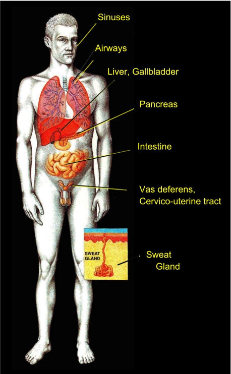
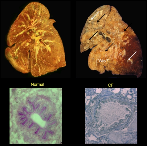
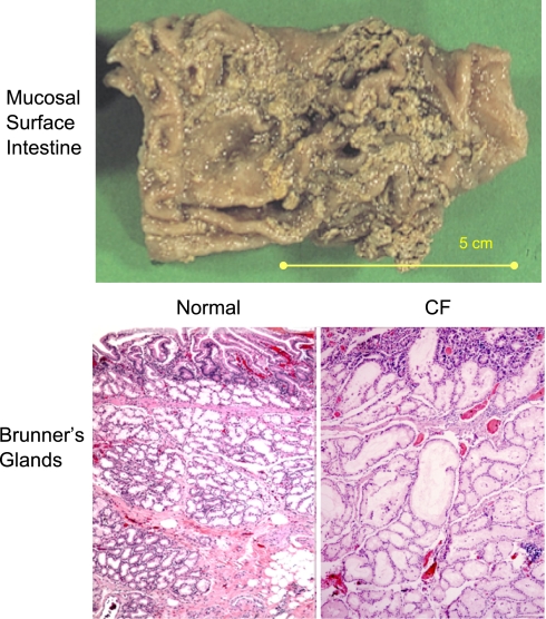
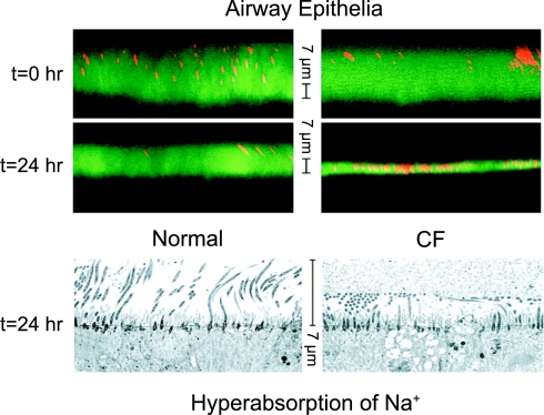
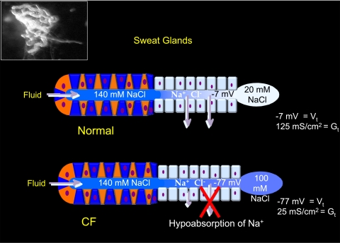
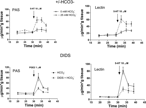
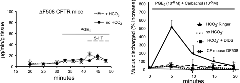
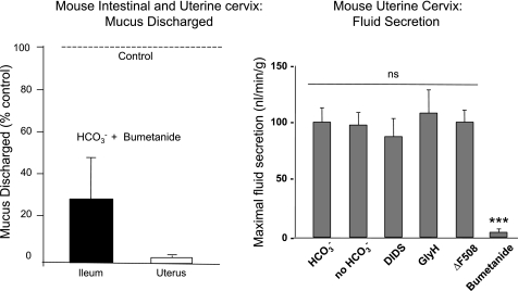
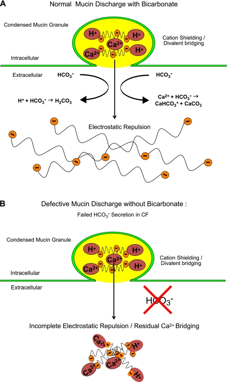
The invitation to present the 2010 Hans Ussing lecture for the Epithelial Transport Group of the American Physiological Society offered me a unique, special, and very surprising opportunity to join in saluting a man whom I met only once, but whose work was the basis, not only for my career, but also for finding the molecular defect in the inherited disease cystic fibrosis (CF). In this context, I will venture to make the tribute with a new explanation of why a mutation in a single gene that codes for an anion channel can cause devastation of multiple epithelial systems with pathogenic mucus. In so doing, I hope to raise awareness of a new role for that peculiar anion around which so much physiology revolves, HCO(3)(-). I begin by introducing CF pathology as I question the name of the disease as well as the prevalent view of the basis of its pathology by considering: 1) mucus, 2) salt, and 3) HCO(3)(-). I then present recent data showing that HCO(3)(-) is required for normal mucus discharge, and I will close with conjecture as to how HCO(3)(-) may support mucus discharge and why the failure to transport this electrolyte is pathogenic in CF.


Figures

Fig. 1.
Fig. 1.
Hans H. Ussing (30 December 1911–22 December 2000). The portrait is from 1955 at the time Ussing became a Fellow of the Royal Danish Academy of Sciences and Letters. Reprinted with permission from the Royal Danish Academy of Sciences and Letters. The drawing depicts what is now known as the “Ussing chamber.” Reprinted from Ussing HH and Zerahn K. Active transport of sodium as the source of electric current in the short-circuited isolated frog skin. Acta Physiol Scand 23: 110–127, 1951, with permission from Wiley.
Fig. 2.
Fig. 2.
A disease of epithelial electrolyte transport. Since much of the pathology is expressed in exocrine systems, Cystic fibrosis could be classified as an exocrinopathy. Inset illustrates an eccrine sweat gland in the skin. Drawing is reprinted with permission from Robert Osti.
Fig. 3.
Fig. 3.
Coronal sections of whole lungs and cross sections of small airways. Note the uniform parenchyma of normal lung (top left) in contrast to the lung from a cystic fibrosis (CF) patient (top right). The latter shows an enormous number of ectatic lesions (arrows) with most airways completely filled and obstructed with purulent mucoid accumulations (black arrows). Examined microscopically, the lumens of normal small airways are virtually free of secretions (bottom left); in contrast, bronchioles from a CF patient can be obstructed and distended by mucus secretions even without significant evidence of inflammation (bottom right). The destruction demands “Why?” a defect in electrolyte transport is so destructive. The coronal sections are courtesies of Walter Finkbeiner, University of California, San Francisco, and the CF cross section is a courtesy of Carl Doershuk (Case-Western Reserve University).
Fig. 4.
Fig. 4.
Mucus adherence and accumulation in CF. Top: a small intestine from a CF patient with mucus accumulated and “stuck” to luminal epithelial surface. Image was kindly provided by Peter Durie and Ernest Cutz, Universty of Toronto, Canada. Bottom: histology of submucosal Brunner's glands of small intestine illustrating normal microanatomy of acini and ducts below the intestinal crypts (bottom left) in contrast to the deranged acini and ducts that have become massively distended by accumulated mucoid secretions (bottom right). Micrographs were provided by Walt Finkbeiner, University of California, San Francisco.
Fig. 5.
Fig. 5.
Prevalent view of mucoviscidosis. Mucus in airway surface fluid is considered to be thick and dessicated in CF due to excessive absorption of Na+ (fluid). Confocal microscopic images in the top show thicknesses of fluid surface layers at time 0 and 24 h later covering cultured normal (left) and CF (right) bronchial epithelial cells. The images below show electron micrographs of tissues 24 h after applying the same fluid load above each culture of cells. The thin CF layer above and the apparently compressed cilia in the image below indicate excessive fluid absorption. Reprinted and adapted from Matsui et al. Evidence for periciliary liquid layer depletion, not abnormal ion composition, in the pathogenesis of cystic fibrosis airways disease. Cell 95: 1005–1015, 1998, with permission from Elsevier.
Fig. 6.
Fig. 6.
Functions of normal and CF sweat glands. Inset: an isolated sweat gland as a single coiled tubule with the secretory coil in the left half of the image connected to, but distinguishable from, the reabsorptive duct in the right half of the image, which is illustrated at the bottom. Thermoregulated sweating is normal in CF and secretes the same load of isotonic precursor sweat from the secretory coil (blue and red cells on the left) to the more distal reabsorptive ducts (pale blue cells on the right). As the secreted fluid passes through the normal duct (top), Na+ and Cl are absorbed in excess of water, yielding a quite hypotonic sweat secretion on the skin surface. In contrast, as the fluid passes through the CF sweat gland, Cl and consequently Na+ cannot be efficiently absorbed, yielding a much higher concentration of excreted salt in the sweat (bottom). Note that even though the rate of salt absorption in the normal duct is fivefold greater than in the CF duct, the transepithelial voltage associated with normal absorption is only about 1/10 of that of the CF duct. Thus the transepithelial potential (Vt) indicates relative separation of charge due to the absorptive process and not the rate of the Na+ absorption.
Fig. 7.
Fig. 7.
HCO3-dependent mucus discharge. Top: lumens of isolated, perfused ileal segments were incubated with 25 mM HCO3 (dashed lines) and without HCO3 (solid lines) in the serosal bathing media. Bottom: luminally perfused segments were incubated in 25 mM HCO3 with (dashed lines) or without DIDS (solid lines) in the serosal media to block HCO3 secretion. Aliquots of perfusates were assayed for mucus content at the times indicated. In either case, interfering with HCO3 secretion reduces mucus discharge. Discharged mucus was assayed by periodic acid Schiff reagent (PAS, left) and confirmed by conjugated WGA-Lectin binding assays (right). Both assays render similar results. Reprinted and adapted with permission from the American Society for Clinical Investigation from Garcia MA, Yang N, and Quinton PM. 2009. Normal mouse intestinal mucus release requires cystic fibrosis transmembrane regulator-dependent bicarbonate secretion. J Clin Invest 119: 2613–2622.
Fig. 8.
Fig. 8.
Mucus discharge in intestine and uterine tracts. Left: intestine from mice homozygous for the ΔF508 mutation show severely blunted response to stimulation by PGE2 (10−6 M) and 5-HT (10−5 M). The amount of mucus release does not differ between segments in the presence or absence of HCO3 when stimulated with PGE2 (n = 5, P = 0.2) or serotonin (5-HT, P = 0.475) assayed by lectin-WGA-DAB. Right: the uterus responds to manipulations of HCO3and HCO3 transport qualitatively in the same manner as does the ileum, but in contrast to the ileum, virtually no mucins appear in the perfusate in the absence of HCO3 or in the presence of the HCO3 transport inhibitor DIDS. As in the intestine, CF tissue does not respond to stimulation even in the presence of HCO3. Reprinted and adapted with permission from the American Society for Clinical Investigation from Garcia MA, Yang N, and Quinton PM. 2009. Normal mouse intestinal mucus release requires cystic fibrosis transmembrane regulator-dependent bicarbonate secretion. J Clin Invest 119: 2613–2622 and adapted from Muchekehu RW and Quinton PM. A new role for bicarbonate secretion in cervico-uterine mucus release. J Physiol 588: 2329–2342, 2010.
Fig. 9.
Fig. 9.
Effects of inhibiting fluid secretion. Adding bumetanide to block Na+/K+/2Cl cotransporter and fluid secretion (right) decreases the amount of mucus discharged (left) into the perfusate of the ileum to <30% and of the uterus to less than a few percent compared with controls without bumetanide (dashed line, 100%) stimulated with PGE2 (ileum) or PGE2 and carbachol (uterus). Fluid secretion measured in the uterus is only blocked by bumetanide (***P < 0.001) and not by the absence of HCO3 or the presence of DIDS, GlyH-101, or the defective gene (ΔF508) expressed in the CF mouse (right). Reprinted and adapted with permission from the American Society for Clinical Investigation from Garcia MA, Yang N, and Quinton PM. 2009. Normal mouse intestinal mucus release requires cystic fibrosis transmembrane regulator-dependent bicarbonate secretion. J Clin Invest 119: 2613–2622 and adapted from Muchekehu RW and Quinton PM. A new role for bicarbonate secretion in cervico-uterine mucus release. J Physiol 588: 2329–2342, 2010.
Fig. 10.
Fig. 10.
Explosion of condensed and compacted mucin. Mucins are stored in granules of goblet cells under conditions of very high Ca2+ concentration and very low pH (top left) to prevent condensed compacted mucin molecules from expanding under the electrostatic forces of their fixed negative charges. Bottom images dramatize mucin expansion as an exploding coiffure that is compacted in “curlers” that explode within seconds into a massive free-flowing mane when exposed to HCO3 as it is discharged; cf. Fig. 11. Top left is from Neutra MR, O'Malley LJ, and Specian RD. Regulation of intestinal goblet cell secretion. II. A survey of potential secretagogues. Am J Physiol Gastrointest Liver Physiol 242: G380–G387, 1982. Bottom left courtesy of Chelsea Johnson-Root; bottom right courtesy of Bayda Bahur.
Fig. 11.
Fig. 11.
Models of effects of HCO3. A: prediction of effects of HCO3 during normal mucin discharge. Repulsive electrostatic forces on the mucin polysaccharides are unshielded as HCO3 competes with the fixed negative sites for Ca2+ and H+, disrupting divalent bridges and forcing the mucin molecules to rapidly expand into an extended network of charged polymers. B: prediction of the effect of poor HCO3 secretion during mucin discharge as might occur in cystic fibrosis. Without competition from HCO3, the repulsive electrostatic forces of the fixed negative sites on the mucin polysaccharides remain shielded and bound by Ca2+ and H+, leaving molecules that adhere to each other and remain relatively condensed and viscous Reprinted and adapted with permission from the American Society for Clinical Investigation from Garcia MA, Yang N, and Quinton PM. 2009. Normal mouse intestinal mucus release requires cystic fibrosis transmembrane regulator-dependent bicarbonate secretion. J Clin Invest 119: 2613–2622.
Fig. 12.
Fig. 12.
Mucoviscidosis in the absence of HCO3. Uterine tissue was incubated in HCO3-free (left) and HCO3-containing (right) Ringer solutions for 20 min and then stimulated with PGE2 + carbachol for 20 min. Significantly more PAS-positive material remains in the lumens of tissue stimulated without HCO3 (arrows, top left) compared with tissue stimulated with HCO3 (top right). Tissue stained for MUC5B mucin (red) and counterstained with DAPI (blue nuclei, bottom) correlates with the PAS staining in the corresponding micrographs above (top: bar 20 μm. bottom: bar 50 μm). Adapted from Muchekehu RW and Quinton PM. A new role for bicarbonate secretion in cervico-uterine mucus release. J Physiol 588: 2329–2342, 2010.

References

    1. Aitken ML, Verdugo P. Donnan mechanism of mucin release and conditioning in goblet cells: the role of polyions. Symp Soc Exp Biol 43: 73–80, 1989 - PubMed
    1. Akiba Y, Guth PH, Engel E, Nastaskin I, Kaunitz JD. Dynamic regulation of mucus gel thickness in rat duodenum. Am J Physiol Gastrointest Liver Physiol 279: G437–G447, 2000 - PubMed
    1. Allen A, Hutton DA, Pearson JP. The MUC2 gene product: a human intestinal mucin. Int J Biochem Cell Biol 30: 797–801, 1998 - PubMed
    1. Andersen DH. Cystic fibrosis of the pancreas and its relation to celiac disease. Am J Dis Children 56: 344–399, 1938
    1. Bijman J, Quinton P. Permeability properties of cell membranes and tight junctions of normal and cystic fibrosis sweat ducts. Pflugers Arch 408: 505–510, 1987 - PubMed

Publication types

MeSH terms