. 2010 Sep 29;5(9):e13074.
doi: 10.1371/journal.pone.0013074.

ERK3 is required for metaphase-anaphase transition in mouse oocyte meiosis

Affiliations

ERK3 is required for metaphase-anaphase transition in mouse oocyte meiosis

Sen Li et al. PLoS One. .

Abstract

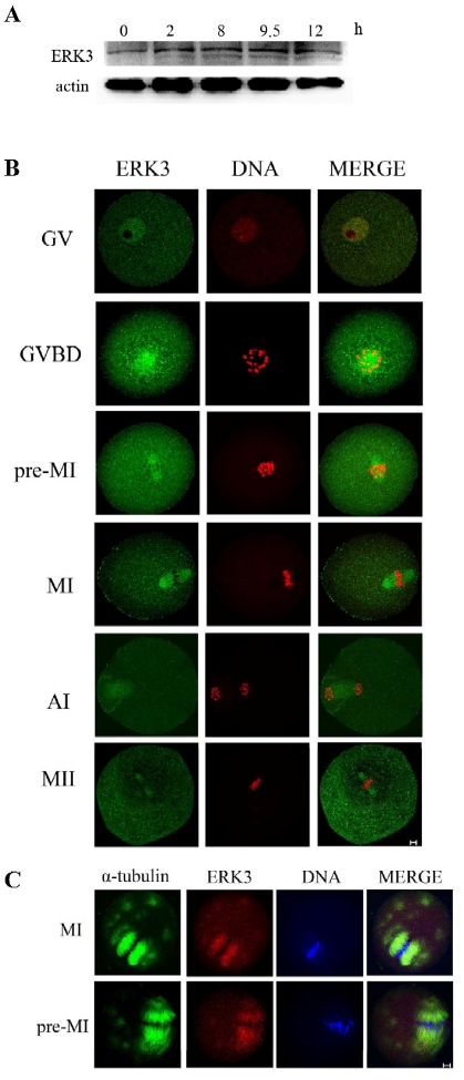
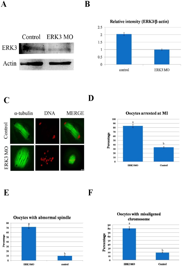
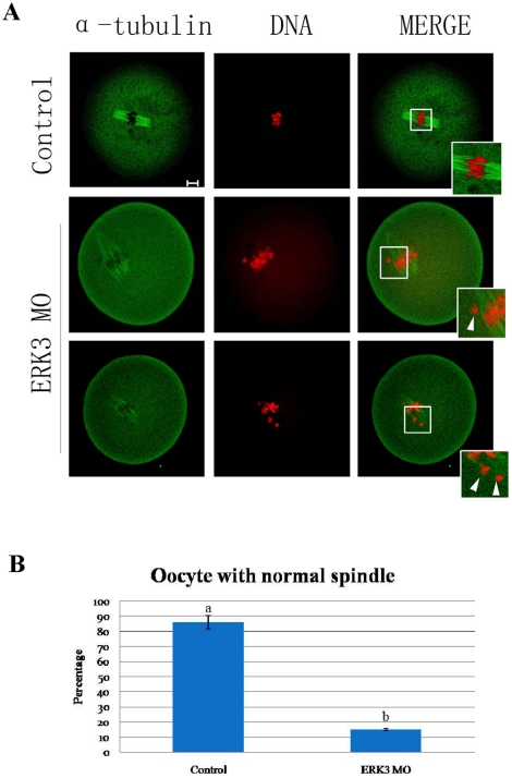
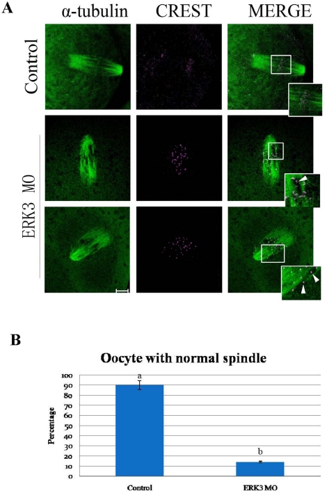
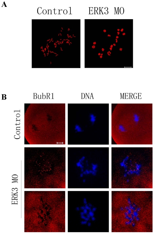
ERK3 (extracellular signal-regulated kinase 3) is an atypical member of the mitogen-activated protein (MAP) kinase family of serine/threonine kinases. Little is known about its function in mitosis, and even less about its roles in mammalian oocyte meiosis. In the present study, we examined the localization, expression and functions of ERK3 during mouse oocyte meiotic maturation. Immunofluorescent analysis showed that ERK3 localized to the spindles from the pre-MI stage to the MII stage. ERK3 co-localized with α-tubulin on the spindle fibers and asters in oocytes after taxol treatment. Deletion of ERK3 by microinjection of ERK3 morpholino (ERK3 MO) resulted in oocyte arrest at the MI stage with severely impaired spindles and misaligned chromosomes. Most importantly, the spindle assembly checkpoint protein BubR1 could be detected on kinetochores even in oocytes cultured for 10 h. Low temperature treatment experiments indicated that ERK3 deletion disrupted kinetochore-microtubule (K-MT) attachments. Chromosome spreading experiments showed that knock-down of ERK3 prevented the segregation of homologous chromosomes. Our data suggest that ERK3 is crucial for spindle stability and required for the metaphase-anaphase transition in mouse oocyte maturation.


Conflict of interest statement

Competing Interests: The authors have declared that no competing interests exist.

Figures

Figure 1
Figure 1. Subcellular localization, expression of ERK3 and localization of ERK3 treated with spindle-perturbing agents.
(A) Samples were collected after oocytes had been cultured for 0.,2, 8, 9.5 and 12 h, corresponding to GV, GVBD, pre-MI, MI,ATI and MII stage, respectively. The molecular mass of ERK3 is 100 kDa and that of β-actin is 42 kDa. (B) Confocal microcopy showing immunostaining of ERK3 (green) and DNA (red) in oocytes at GV, GVBD, pro-M I, M I, A I and M II stages. (C) Oocytes at the metaphase I stage were incubated in M2 medium containing 10 µM taxol for 45 minutes and then double stained with antibodies against ERK3 as well as α-tubulin. Green, α-tubulin; red, ERK3; blue, DNA; yellow, overlapping of green and red. Each sample was counterstained with Hoechst 33258 to visualize DNA. Bar = 10 µm.
Figure 2
Figure 2. ERK3 deletion arrested oocytes at the MI stage and led to decreased spindle stability.
After microinjection of ERK3 MO, the oocytes were incubated in M2 medium containing 2.5 µM milrinone for 21 h, then transferred to milrinone-free M2 medium for 10 h. (A) Western blot of ERK3 in the ERK3 MO group and control group. The ERK3 molecular mass is 100 kDa and that of actin is 42 kDa. (B) Relative intensity of ERK3/β-actin was assessed by volume analysis. (C) After microinjection, oocytes microinjected with ERK3 morpholino were arrested at the MI stage at 10 h of culture, but the control oocytes were in the AI stage. Double staining of α-tubulin (green) and DNA (red). Bar = 10 µm. (D) Percentage of oocytes in the ERK3 MO microinjected group (n = 42) and control group (n = 40). Data are presented as mean ± SE. Different superscripts indicate statistical difference (p<0.05). (E) Percentage of oocytes with abnormal spindles in the ERK3 MO injected group (n = 45) and control group (n = 44). (F) Percentage of oocytes with misaligened chromosomes in the ERK3 MO injected group (n = 45) and control morpholino injected group (n = 44). Data are presented as mean ± SE. Different superscripts indicate statistical difference (p<0.05).
Figure 3
Figure 3. Deletion of ERK3 induced unstable microtubule-chromosome attachments at the MI stage.
(A) Oocytes of control and ERK3 MO groups were cultured for 8.5 h followed by cold treatment for 10 minutes in M2 medium which was pre-cooled at 4°C. Magnifications of the boxed regions show that all chromosomes were attached to microtubules in control oocytes, but not in ERK3 MO microinjected oocytes, whereas abnormal spindles were observed in ERK3 MO group. Bar = 10 µm. (B) Percentage of oocytes with normal spindles in the ERK3 MO group (n = 50) and control group (n = 49). Data are presented as mean ± SE. Different superscripts indicate statistical difference (p<0.05). Bar = 10 µm.
Figure 4
Figure 4. ERK3 deletion disrupted the attachments between kinetochores and microtubules.
(A) Oocytes of control and ERK3 MO groups were cultured for 8.5 h followed by cold treatment for 10 minutes in M2 medium which was pre-cooled at 4°C. Magnifications of the boxed regions showed intact attachments between kinetochores and microtubules in the control group, but not in the ERK3 MO group, whereas abnormal spindles were observed in the ERK3 MO group. Bar = 10 µm. (B) Percentage of oocytes with normal spindles in the control group (52) and the ERK3 MO group (51). Data are presented as mean ± SE. Different superscripts indicate statistical difference (p<0.05).
Figure 5
Figure 5. ERK3 deletion inhibited chromosome segregation and activated SAC protein BubR1.
(A) Oocytes of control and ERK3 MO groups were cultured for 10 h, followed by chromosome spreading experiments. (B) Detection of BubR1 in oocytes in control and ERK3 MO groups. Red, BubR1; blue, DNA. Bar = 10 µm.

References

    1. Hassold T, Hunt P. To err (meiotically) is human: the genesis of human aneuploidy. Nat Rev Genet. 2001;2:280–291. - PubMed
    1. Mehlmann LM. Stops and starts in mammalian oocytes: recent advances in understanding the regulation of meiotic arrest and oocyte maturation. Reproduction. 2005;130:791–799. - PubMed
    1. Sirard MA. Resumption of meiosis: mechanism involved in meiotic progression and its relation with developmental competence. Theriogenology. 2001;55:1241–1254. - PubMed
    1. Doubilet S, McKim KS. Spindle assembly in the oocytes of mouse and Drosophila--similar solutions to a problem. Chromosome Res. 2007;15:681–696. - PubMed
    1. Musacchio A, Salmon ED. The spindle-assembly checkpoint in space and time. Nat Rev Mol Cell Biol. 2007;8:379–393. - PubMed

Publication types

Substances