

Novel Gd nanoparticles enhance vascular contrast for high-resolution magnetic resonance imaging
- PMID: 20927340
- PMCID: PMC2948015
- DOI: 10.1371/journal.pone.0013082
Novel Gd nanoparticles enhance vascular contrast for high-resolution magnetic resonance imaging
Abstract
Background: Gadolinium (Gd), with its 7 unpaired electrons in 4f orbitals that provide a very large magnetic moment, is proven to be among the best agents for contrast enhanced MRI. Unfortunately, the most potent MR contrast agent based on Gd requires relatively high doses of Gd. The Gd-chelated to diethylene-triamine-penta-acetic acid (DTPA), or other derivatives (at 0.1 mmole/kg recommended dose), distribute broadly into tissues and clear through the kidney. These contrast agents carry the risk of Nephrogenic Systemic Fibrosis (NSF), particularly in kidney impaired subjects. Thus, Gd contrast agents that produce higher resolution images using a much lower Gd dose could address both imaging sensitivity and Gd safety.
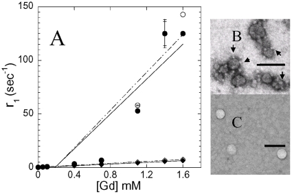
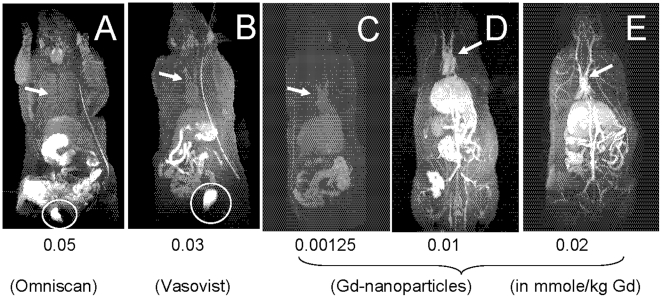
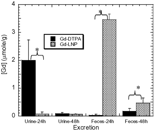
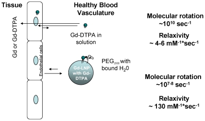
Methodology/principal findings: To determine whether a biocompatible lipid nanoparticle with surface bound Gd can improve MRI contrast sensitivity, we constructed Gd-lipid nanoparticles (Gd-LNP) containing lipid bound DTPA and Gd. The Gd-LNP were intravenously administered to rats and MR images collected. We found that Gd in Gd-LNP produced a greater than 33-fold higher longitudinal (T(1)) relaxivity, r(1), constant than the current FDA approved Gd-chelated contrast agents. Intravenous administration of these Gd-LNP at only 3% of the recommended clinical Gd dose produced MRI signal-to-noise ratios of greater than 300 in all vasculatures. Unlike current Gd contrast agents, these Gd-LNP stably retained Gd in normal vasculature, and are eliminated predominately through the biliary, instead of the renal system. Gd-LNP did not appear to accumulate in the liver or kidney, and was eliminated completely within 24 hrs.
Conclusions/significance: The novel Gd-nanoparticles provide high quality contrast enhanced vascular MRI at 97% reduced dose of Gd and do not rely on renal clearance. This new agent is likely to be suitable for patients exhibiting varying degrees of renal impairment. The simple and adaptive nanoparticle design could accommodate ligand or receptor coating for drug delivery optimization and in vivo drug-target definition in system biology profiling, increasing the margin of safety in treatment of cancers and other diseases.
Conflict of interest statement
Figures
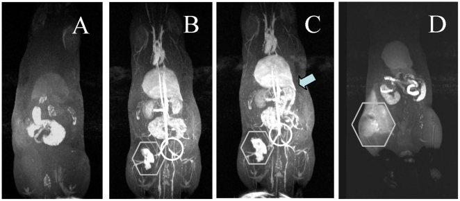
 at the left femoral vein, Gd localization is apparent as a high contrast area. Please note the vaso-restriction ○ clearly apparent in panels B & C.
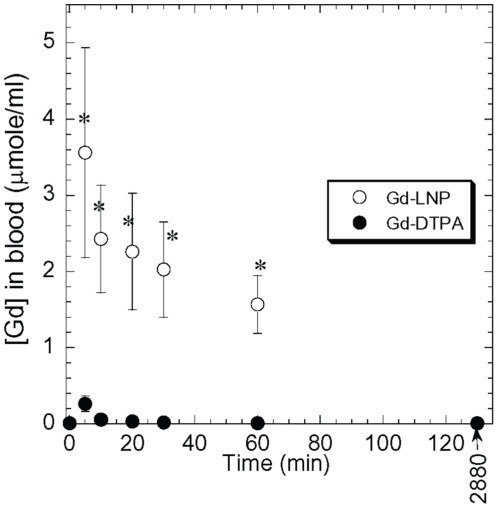
References
-
- Rofsky NM, Sherry AD, Lenkinski RE. Nephrogenic systemic fibrosis: a chemical perspective. Radiology. 2008;247:608–612. - PubMed
-
- FDA. 2007. Information for Healthcare Professionals Gadolinium-Based Contrast Agents for Magnetic Resonance Imaging (marketed as Magnevist, MultiHance, Omniscan, OptiMARK, ProHance). FDA ALERT: US FDA. This updated Alert highlights FDA's request for addition of a boxed warning and new warnings about risk of nephrogenic systemic fibrosis (NSF) to the full prescribing information for all gadoliniumbased contrast agents (GBCAs) (Magnevist, MultiHance, Omniscan, OptiMARK, ProHance)
-
- Huang SK, Lee KD, Hong K, Friend DS, Papahadjopoulos D. Microscopic localization of sterically stabilized liposomes in colon carcinoma-bearing mice. Cancer Res. 1992;52:5135–5143. - PubMed
-
- Hak S, Sanders HM, Agrawal P, Langereis S, Grull H, et al. A high relaxivity Gd(III)DOTA-DSPE-based liposomal contrast agent for magnetic resonance imaging. Eur J Pharm Biopharm 2008 - PubMed
-
- Meaney JF, Goyen M. Recent advances in contrast-enhanced magnetic resonance angiography. Eur Radiol. 2007;17(Suppl 2):B2–6. - PubMed
Publication types
MeSH terms
Substances
Grants and funding
LinkOut - more resources
Full Text Sources
Other Literature Sources
