Review
. 2011 Jan;68(2):243-74.
doi: 10.1007/s00018-010-0536-y. Epub 2010 Oct 8.

The gap gene network

Affiliations
Review

The gap gene network

Johannes Jaeger. Cell Mol Life Sci. 2011 Jan.

Abstract

Gap genes are involved in segment determination during the early development of the fruit fly Drosophila melanogaster as well as in other insects. This review attempts to synthesize the current knowledge of the gap gene network through a comprehensive survey of the experimental literature. I focus on genetic and molecular evidence, which provides us with an almost-complete picture of the regulatory interactions responsible for trunk gap gene expression. I discuss the regulatory mechanisms involved, and highlight the remaining ambiguities and gaps in the evidence. This is followed by a brief discussion of molecular regulatory mechanisms for transcriptional regulation, as well as precision and size-regulation provided by the system. Finally, I discuss evidence on the evolution of gap gene expression from species other than Drosophila. My survey concludes that studies of the gap gene system continue to reveal interesting and important new insights into the role of gene regulatory networks in development and evolution.


Figures

Fig. 1
Fig. 1
Segment determination in Drosophila. a The first 3 h of development of Drosophila melanogaster. Numbers indicate cleavage cycle number, where cycle n covers the time between mitosis n − 1 and mitosis n. The blastoderm stage lasts from 1 min into cycle 10 to the onset of gastrulation (grey background). The embryo remains syncytial (without membranes between nuclei) until cellularization occurs during cycle 14A. The cellular blastoderm stage is more or less instantaneous, since gastrulation begins immediately after cellularization is complete. Cycle 14B denotes the part of cycle 14, which occurs after the onset of gastrulation. Embryos are shown with the anterior pole to the top. b The regulatory hierarchy of the Drosophila segmentation gene network. Segment determination is based on a molecular pre-pattern established by the segmentation genes, which are active during the blastoderm stage. Different regulatory tiers of the network can be distinguished based on mutant phenotypes, epistatic interactions, and expression patterns. Maternal co-ordinate genes are expressed in broad gradients (Bcd protein distribution is shown as an example). They regulate the zygotic gap genes, expressed in broad overlapping domains (the central domain of Kr is shown). Gap genes and pair-rule genes together regulate pair-rule genes, which are expressed in 7–8 stripes (shown for Even-skipped (Eve) protein). Pair-rule genes in turn regulate segment-polarity genes whose expression in 14 stripes becomes established just before the onset of gastrulation (shown for en mRNA). These stripes constitute the segmentation pre-pattern and correspond to the positions of parasegmental boundaries later in development. Arrows indicate regulatory interactions between classes of segmentation genes. Circular arrows represent cross-regulation within a class. Embryo images are shown with anterior to the left, and dorsal up (see text for details). a is reproduced with permission from the Journal of Cell Science: http://jcs.biologists.org [30]. b Embryo images (Bcd, Kr, and Eve) are from the FlyEx database [164, 166]. The image of en is courtesy of Carlos E. Vanario-Alonso
Fig. 2
Fig. 2
Maternal gradients and French Flags. ac Three maternal systems regulate the expression of gap genes: a The anterior system is based on the Bcd gradient, which regulates gap gene transcription in a concentration-dependent manner and also establishes the posterior gradient of Cad through translational repression. b The posterior system is based on the Nos gradient, whose only function is to repress the translation of maternal hb mRNA in the posterior region of the embryo to form an anterior Hb protein gradient. c The terminal system is based on Tor signaling from both terminal ends of the embryo, which induces the expression of the terminal gap genes tll and hkb at both poles of the embryo. Expression profiles are based on integrated data from the FlyEx database [164, 166], except for Nos, which is illustrated by a mirrored Bcd gradient due to the absence of quantitative Nos expression data. d Wolpert’s French Flag model: A morphogen is produced at a source (shown in green), diffuses through the tissue (without protein degradation) and is degraded at a sink (pink), at the other end of the tissue. Specific concentration thresholds in the resulting linear gradient (T 1, T 2) are detected by cells (or nuclei) in the tissue, which switch on alternative target genes (represented by blue, white, and red), which in turn lead to distinct differentiation pathways in each region of the embryo. In this model, development is seen as a two-step process: First, positional information is implemented by the morphogen gradient (step 1). Subsequently, cells in the tissue passively interpret this information (step 2). Concentration thresholds in the gradient correspond exactly to borders of downstream expression territories. e A revised French Flag, incorporating target domain shifts and increasing precision over time. New evidence shows that maternal gradients are not sufficient to determine precise downstream boundary positions on their own. Instead, cross-regulation among target genes leads to (a) shifts in boundary positions over time and (b) an observed increase in the precision with which boundaries are placed. In this model, there is no longer a precise correspondence between concentration thresholds in the gradient and the final position of target domain boundaries
Fig. 3
Fig. 3
Early versus late gap gene regulation. Gap gene regulation can be divided into two distinct phases: early regulation of gap mRNA domains is based on maternal gradients only, while late regulation of protein domains involves gap–gap cross-regulatory interactions. The position of gap domains along the major, or antero-posterior (A–P) axis of the embryo is shown schematically as colored boxes. Only the trunk region of the embryo (approx. 35–95% A–P position) is included in the diagram. Anterior is to the left, posterior to the right. Background color represents activating inputs by Bcd and Cad. Top panel: arrowheads represent activating; T-bars represent repressive inputs responsible for setting specific domain boundaries. Bottom panel: arrows and T-bars represent activating and repressive gap–gap cross-regulation, respectively. Circular arrows represent auto-activation. The thickness of the T-bars corresponds to repressive strength. Question marks indicate missing or ambiguous evidence, or other open questions regarding gap gene regulation (see text for details)
Fig. 4
Fig. 4
Early gap gene expression. mRNA distribution is visualized by fluorescent in situ hybridization for Kr, kni, and gt during early blastoderm stage (cycles 11–13). The inset shows transient early Kr expression during mitosis 11. Embryo images are from [19], shown with anterior to the left, dorsal up. Plots show individual one-dimensional expression profiles for each gene from the middle 10% along the dorso-ventral (D–V) axis at late cycle 13, illustrating the large embryo-to-embryo variability of the patterns at this stage. Relative mRNA concentration is plotted against position along the A–P axis (in %, where 0% is the anterior pole) (see [19, 165] for details on data quantification)
Fig. 5
Fig. 5
Late gap gene expression showing dynamic shifts in gap domain positions. Protein expression patterns are shown for Hb, Kr, Kni, and Hb at eight time classes during cycle 14A (T1–T8) [61]. Plots show integrated one-dimensional expression patterns from the middle 10% along the D–V axis over time, illustrating the anterior shift in boundary position for all expression domains posterior of the central Kr domain. Relative protein concentration is plotted against position along the A–P axis (in %, where 0% is the anterior pole). Embryo images and integrated data for plots are from the FlyEx database [164, 166], shown with anterior to the left, dorsal up (see [165] for details on data quantification)
Fig. 6
Fig. 6
Two ways of setting expression domain boundaries. Such boundaries can only be set by an activation threshold (left)—which implies the same polarity for the regulator gradient and the regulated boundary—or by repression (right)—which implies opposite polarity for regulator and regulated target
Fig. 7
Fig. 7
The five main regulatory mechanisms for late gap gene regulation: a Gap genes are activated by maternal Bcd and Cad in broad regions of the embryo. b Auto-activation leads to intensification and sharpening of domain boundaries in specific gap domains. c Strong cross-repression between gap genes with mutually exclusive expression domains leads to the basic staggered arrangement of gap domains (alternating cushions hypothesis). d Weaker cross-repression between gap genes with overlapping domains of expression leads to anterior shifts in boundary positions over time. e Repression by terminal gap genes establishes the posterior boundaries of several gap domains and excludes gap gene expression from the un-segmented terminal regions of the embryo. Horizontal axis, background color, gap domains, and regulatory links as in Fig. 3. Colored picture elements highlight those domains involved in or affected by a specific mechanism
Fig. 8
Fig. 8
Molecular mechanisms of gap gene regulation. Transcripts (start site is indicated by arrow, exons by grey boxes, and introns by thin triangular lines) and protein coding sequence (black boxes), as well as cis-regulatory elements (CREs; thick black bars) involved in gap gene regulation are shown schematically for hb (a), Kr (b), kni (c), and gt (d). Solid and dashed curved arrows in a indicate early regulation by separate CREs and late regulation by a common CRE, respectively. Inset in b shows repression by competitive binding, c shows repression by interactions between CREs (kni_kd is composed of 223 and 64 bp-sub-elements; Hb-binding to the 223-bp element masks Bcd-activation in the 64-bp element in the posterior of the embryo), and d shows that strong repression of gt by Hb (required for the anterior boundary of the posterior domain) must be overcome for correct expression in the anterior domain. Genomic positions are not drawn to scale (see text for details)
Fig. 9
Fig. 9
The evolution of the gap gene system. a A simplified phylogenetic tree for the arthropods is shown to the left (based on [323, 360, 361]) indicating relationships between taxa containing species in which gap genes have been studied in some detail. The prevalent mode of segment determination is shown in the first column (S short-, L long-germband). The presence or absence of an oscillator based on Notch-signaling is indicated in the second column. Evidence for or against gap-like expression patterns and phenotypes for the trunk gap genes hb, Kr, kni, and gt is indicated in the remaining two columns to the right (see key for abbreviations). b A simplified phylogenetic tree for the diptera (based on [362]) is shown to the left, indicating relationships between dipteran families containing species in which gap genes have been studied in some detail. The presence or absence of maternal gradients is indicated in the first column (see key for abbreviations). Only higher (cyclorrhaphan) flies have a Bcd gradient. The relative position of gap domains [from left to right in Drosophila: gt, hb (anterior), Kr, kni, gt, and hb (posterior)] and the number of pair-rule (eve) stripes before gastrulation are shown schematically to the right. There are two convergent branches, which have evolved an extreme form of long-germband development: Mosquitoes (Culicidae, top) and higher flies (Phoridae, Syrphidae, Tephritidae, and Drosophilidae, bottom) show seven eve stripes and posterior gt/hb domains before gastrulation. In contrast, midges (Psychodidae/Scatopsidae) lack posterior gt/hb and only develop 3–6 eve stripes during the blastoderm stage. Note that the posterior domains of hb and gt have swapped positions (double arrow) in mosquitoes. Question marks indicate unknown gap gene expression patterns (see main text for details)

References

    1. Akam M. The molecular basis for metameric pattern in the Drosophila embryo. Development. 1987;101:1–22. - PubMed
    1. Ingham PW. The molecular genetics of embryonic pattern formation in Drosophila . Nature. 1988;335:25–34. - PubMed
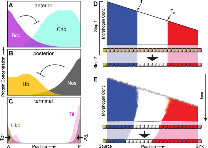
    1. Wolpert L. The French Flag problem: a contribution to the discussion on pattern development and regulation. In: Waddington CH, editor. Towards a theoretical biology. Edinburgh, UK: Edinburgh University Press; 1968. pp. 125–133.
    1. Wolpert L. Positional information and the spatial pattern of cellular differentiation. J Theor Biol. 1969;25:1–47. - PubMed
    1. Jaeger J, Reinitz J. On the dynamic nature of positional information. BioEssays. 2006;28:1102–1111. - PubMed

MeSH terms

Substances

LinkOut - more resources