Review
. 2010 Oct 7;12(1):55.
doi: 10.1186/1532-429X-12-55.

Cardiovascular magnetic resonance at 3.0 T: current state of the art

Affiliations
Review

Cardiovascular magnetic resonance at 3.0 T: current state of the art

John N Oshinski et al. J Cardiovasc Magn Reson. .

Abstract

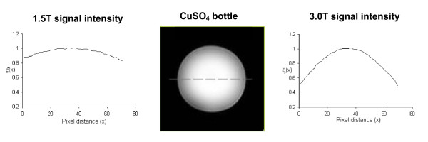
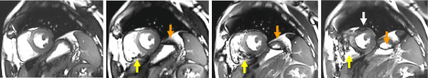
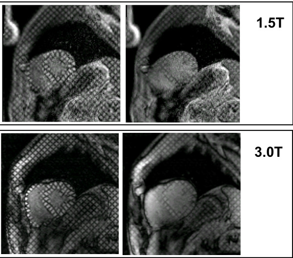
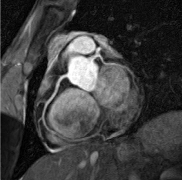
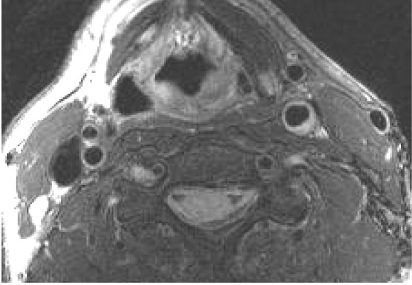
There are advantages to conducting cardiovascular magnetic resonance (CMR) studies at a field strength of 3.0 Telsa, including the increase in bulk magnetization, the increase in frequency separation of off-resonance spins, and the increase in T1 of many tissues. However, there are significant challenges to routinely performing CMR at 3.0 T, including the reduction in main magnetic field homogeneity, the increase in RF power deposition, and the increase in susceptibility-based artifacts.In this review, we outline the underlying physical effects that occur when imaging at higher fields, examine the practical results these effects have on the CMR applications, and examine methods used to compensate for these effects. Specifically, we will review cine imaging, MR coronary angiography, myocardial perfusion imaging, late gadolinium enhancement, and vascular wall imaging.


Figures

Figure 1
Figure 1
The field-focusing effect is illustrated in a phantom image. Relative signal intensity profiles (ξ) are shown as a function of position (x) in a CuSO4 bottle. On the left is a signal intensity profile for a 90° flip angle in a 1.5T scanner. Signal intensity is fairly uniform across the bottle. On the right is a signal intensity profile for a 90° flip angle in a 3.0T scanner. Signal intensity is focused at the center of the bottle.
Figure 2
Figure 2
Short-axis, SSFP images acquired at four different repetition times (TR's), ranging from 2.3msec (far left) to 5.0msec (far right). As TR is increased, artifacts due to susceptibility and field inhomogeneity are seen in the RV (yellow arrow), at the diaphragm (orange arrow), is and of the anterior and lateral walls of myocardium (white arrows).
Figure 3
Figure 3
Tagging images acquired in systole and mid-diastole at 3.0T and 1.5T. Note that in diastole tag lines have faded more at 1.5T than at 3.0T. The reduced fading at 3.0T is due to the prolonged T1 at 3.0T. The longer lasting tag lines may allow for better analysis of diastolic function at 3.0T. Signal-to-noise is also higher in the 3.0T images.
Figure 4
Figure 4
Image of the coronary arteries obtained at 3.0T using a 32-channel coil and employing a targeted 3 D GRE approach. Pixel resolution is 0.7 × 1.0 × 2.0. The right coronary artery (RCA) and left circumflex artery (LCX) and branching vessels can easily be seen. In general, the 3.0T coronary images have superior SNR and CNR compared to 1.5T.
Figure 5
Figure 5
Image of the carotid artery obtained in a subject with a thickened wall in the left common carotid artery. The image was acquired at 3.0T with a four-channel carotid coil. Image quality as assessed by SNR and CNR is superior to 1.5T images

References

    1. Alvarez-Linera J. 3T MRI: advances in brain imaging. Eur J Radiol. 2008;67(3):415–26. doi: 10.1016/j.ejrad.2008.02.045. - DOI - PubMed
    1. DeLano MC, Fisher C. 3T MR imaging of the brain. Magn Reson Imaging Clin N Am. 2006;14(1):77–88. doi: 10.1016/j.mric.2006.01.004. - DOI - PubMed
    1. Frayne R, Goodyear BG, Dickhoff P, Lauzon ML, Sevick RJ. Magnetic resonance imaging at 3.0 Tesla: challenges and advantages in clinical neurological imaging. Investigative radiology. 2003;38(7):385–402. doi: 10.1097/00004424-200307000-00003. - DOI - PubMed
    1. Gharib AM, Elagha A, Pettigrew RI. Cardiac magnetic resonance at high field: promises and problems. Curr Probl Diagn Radiol. 2008;37(2):49–56. doi: 10.1067/j.cpradiol.2007.11.003. - DOI - PMC - PubMed
    1. Lee VS, Hecht EM, Taouli B, Chen Q, Prince K, Oesingmann N. Body and cardiovascular MR imaging at 3.0 T. Radiology. 2007;244(3):692–705. doi: 10.1148/radiol.2443060582. - DOI - PubMed

Publication types