. 2010 Dec 3;285(49):38093-103.
doi: 10.1074/jbc.M110.162388. Epub 2010 Oct 7.

Interactions of STAP-2 with Brk and STAT3 participate in cell growth of human breast cancer cells

Affiliations

Interactions of STAP-2 with Brk and STAT3 participate in cell growth of human breast cancer cells

Osamu Ikeda et al. J Biol Chem. .

Abstract

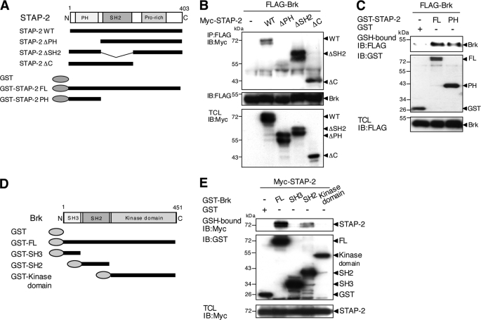
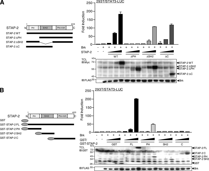
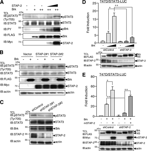
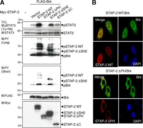
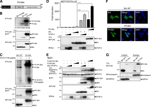
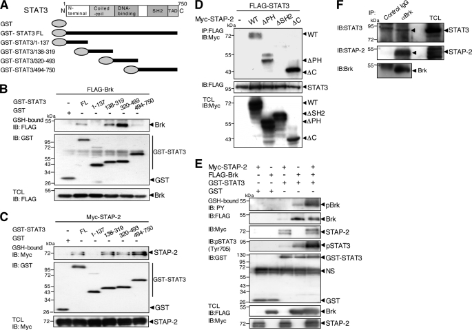
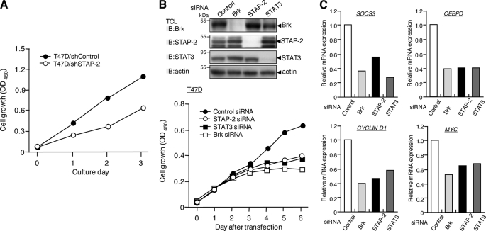
STAP-2 (signal transducing adaptor protein-2) is a recently identified adaptor protein that contains pleckstrin homology (PH) and Src homology 2-like domains, as well as a STAT3-binding motif in its C-terminal region. STAP-2 is also a substrate of breast tumor kinase (Brk). In breast cancers, Brk expression is deregulated and promotes STAT3-dependent cell proliferation. In the present study, manipulated STAP-2 expression demonstrated essential roles of STAP-2 in Brk-mediated STAT3 activation. STAP-2 interacts with both Brk and STAT3. In addition, small interfering RNA-mediated reduction of endogenous STAP-2 expression strongly decreased Brk-mediated STAT3 activation in T47D breast cancer cells. The PH domain of STAP-2 is involved in multiple steps: the binding between Brk and STAP-2, the activation and tyrosine phosphorylation of STAT3, and the activation of Brk. Notably, a STAP-2 PH-Brk fusion protein exhibited robust kinase activity and increased activation and tyrosine phosphorylation of STAT3. Finally, STAP-2 knockdown in T47D cells induced a significant decrease of proliferation, as strong as that of Brk or STAT3 knockdown. Taken together, our findings are likely to inform the development of a novel therapeutic strategy, as well as the determination of novel prognostic values, in breast carcinomas.


Figures

FIGURE 1.
FIGURE 1.
STAP-2 regulates Brk-mediated STAT3 activation in breast cancer cells. A, MCF7 cells in a 24-well plate were transfected with STAT3-LUC (100 ng) and/or FLAG-tagged Brk (100 ng) and expression vector for Myc-tagged STAP-2 WT (30 or 300 ng). At 48 h after transfection, the cells were harvested, and the luciferase activities were measured. At least three independent experiments were carried out for each assay. The error bars represent the S.D. **, p < 0.01. An aliquot of each total cell lysate (TCL) was analyzed by immunoblotting (IB) with an anti-FLAG or anti-Myc antibody. B, T47D cells in a 24-well plate were transfected with STAT3-LUC (100 ng) and/or FLAG-tagged Brk (100 ng) and expression vector for Myc-tagged STAP-2 WT (30 or 300 ng). At 48 h after transfection, the cells were harvested, and the luciferase activities were measured. At least three independent experiments were carried out for each assay. The error bars represent the S.D. **, p < 0.01. An aliquot of each TCL was analyzed by immunoblotting with an anti-FLAG or anti-Myc antibody. C, T47D cells in a 24-well plate were transfected with control or STAP-2 (#1 and #2) siRNA, and the cells were then transfected with STAT3-LUC (100 ng) and/or FLAG-tagged Brk (300 ng) using jetPEI. At 36 h after transfection, the cells were harvested and assayed for luciferase activity using a dual luciferase reporter assay system. The results are indicated as fold induction of luciferase activity from triplicate experiments, and the error bars represent the S.D. **, p < 0.01. An aliquot of each TCL was analyzed by immunoblotting with an anti-Brk, anti-STAP-2, or anti-actin antibody.
FIGURE 2.
FIGURE 2.
Molecular interactions between STAP-2 and Brk. A, schematic diagrams of the domain structures of the STAP-2 deletion mutant fragments are shown. B, 293T cells (1 × 10⁷) were transfected with FLAG-tagged Brk (10 μg) with or without Myc-tagged STAP-2 deletion mutants (8 μg). At 48 h after transfection, the cells were lysed, immunoprecipitated (IP) with an anti-FLAG antibody, and immunoblotted (IB) with an anti-Myc or anti-FLAG antibody. An aliquot of each TCL was immunoblotted with an anti-Myc antibody. C, 293T cells (1 × 10⁷) were transfected with FLAG-tagged Brk (10 μg) with or without GST, GST-fused STAP-2 WT, or PH (8 μg). At 48 h after transfection, the cells were lysed, pulled down with glutathione-Sepharose, and immunoblotted with an anti-FLAG or anti-GST antibody. An aliquot of each TCL was immunoblotted with an anti-FLAG antibody. D, schematic diagrams of the domain structures of the GST-fused Brk deletion mutant fragments are shown. E, 293T cells (1 × 10⁷) were transfected with Myc-tagged STAP-2 (8 μg) with or without GST-fused Brk deletion mutants (10 μg). At 48 h after transfection, the cells were lysed, pulled down with glutathione-Sepharose, and immunoblotted with an anti-Myc or anti-GST antibody. An aliquot of each TCL was immunoblotted with an anti-Myc antibody.
FIGURE 3.
FIGURE 3.
Functional role of the PH domain of STAP-2 in Brk-mediated STAT3 activation. A, schematic diagrams of the domain structures of the STAP-2 deletion mutant fragments are shown. 293T cells in a 24-well plate were transfected with STAT3-LUC (200 ng) and/or FLAG-tagged Brk (100 ng) and increasing amounts (1.0, 10, and 100 ng) of expression vector for Myc-tagged STAP-2 WT, STAP-2 ΔPH, STAP-2 ΔSH2, or STAP-2 C. At 48 h transfection, the cells were harvested, and the luciferase activities were measured. At least three independent experiments were carried out for each assay. The error bars represent the S.D. An aliquot of each TCL was analyzed by immunoblotting (IB) with an anti-Myc or anti-FLAG antibody. B, schematic diagrams of the domain structures of the GST-fused STAP-2 deletion mutant fragments are shown. 293T cells in a 24-well plate were transfected with STAT3-LUC (200 ng) and/or FLAG-tagged Brk (100 ng) and increasing amounts (1, 10, and 100 ng) of expression vector for GST or GST-fused STAP-2 FL, STAP-2 PH, STAP-2 SH2, or STAP-2 C. At 48 h after transfection, the cells were harvested, and the luciferase activities were measured. At least three independent experiments were carried out for each assay. The error bars represent the S.D. An aliquot of each TCL was analyzed by immunoblotting with an anti-GST or anti-FLAG antibody.
FIGURE 4.
FIGURE 4.
STAP-2 regulates Brk-induced tyrosine phosphorylation of STAT3 at Tyr-705. A, MCF7 cells in a 12-well plate were transfected with or without Myc-tagged STAP-2 WT (0.3 and 1.0 μg) and/or FLAG-tagged Brk (+, 0.3 μg; ++, 1.0 μg). At 48 h after transfection, the cells were lysed and immunoblotted (IB) with an anti-pSTAT3 (Tyr-705), anti-STAT3, anti-PY, anti-FLAG, or anti-Myc antibody. B, MCF7/pcDNA3, MCF7/STAP-2#1, and MCF7/STAP-2#2 cells in a 12-well plate were transfected with or without FLAG-tagged Brk (1.0 μg). At 48 h after transfection, the cells were lysed and immunoblotted with an anti-pSTAT3 (Tyr-705), anti-STAT3, anti-FLAG, anti-Myc, or anti-actin antibody. C, T47D/shControl, T47D/shSTAP-2#1, and T47D/shSTAP-2#2 cells were lysed and immunoblotted with an anti-pSTAT3 (Tyr-705), anti-STAT3, anti-Brk, anti-STAP-2, or anti-actin antibody. D, T47D/shControl or T47D/shSTAP-2#1 cells in a 24-well plate were transfected with STAT3-LUC (100 ng) and/or FLAG-tagged Brk (100 or 300 ng). At 48 h after transfection, the cells were harvested, and the luciferase activities were measured. At least three independent experiments were carried out for each assay. The error bars represent the S.D. *, p < 0.05; **, p < 0.01. An aliquot of each TCL was analyzed by immunoblotting with an anti-FLAG, anti-STAP-2, or anti-actin antibody. E, T47D/shControl and T47D/shSTAP-2#1 cells in a 24-well plate were transfected with STAT3-LUC (100 ng) and/or FLAG-tagged Brk (300 ng) and/or expression vector for Myc-tagged mouse STAP-2 WT. At 48 h after transfection, the cells were harvested, and the luciferase activities were measured. At least three independent experiments were carried out for each assay. The error bars represent the S.D. *, p < 0.05; **, p < 0.01. An aliquot of each TCL was analyzed by immunoblotting with an anti-FLAG, anti-Myc, anti-STAP-2, or anti-actin antibody.
FIGURE 5.
FIGURE 5.
STAP-2 PH domain regulates Brk-induced tyrosine phosphorylation of STAT3 at Tyr-705 and nuclear co-localization with Brk. A, 293T cells in a 12-well plate were transfected with FLAG-tagged Brk (1.0 μg) with or without Myc-tagged STAP-2 deletion mutants (1.0 μg). At 48 h after transfection, the cells were lysed and immunoblotted (IB) with an anti-pSTAT3 (Tyr-705), anti-STAT3, anti-PY (short exposure and long exposure for ECL detection), anti-FLAG, or anti-Myc antibody. B, HeLa cells in 12-well plates were transfected with FLAG-tagged Brk (1.0 μg), together with Myc-tagged STAP-2, or Myc-tagged STAP-2 ΔPH (1.0 μg) using jetPEI. At 48 h after transfection, the cells were fixed, incubated with anti-FLAG or anti-Myc antibodies, and visualized with FITC- and rhodamine-conjugated secondary antibodies. The same slides were also stained with DAPI to detect the nuclei.
FIGURE 6.
FIGURE 6.
A STAP2 PH-Brk fusion protein exhibited robust kinase activity and increased activation and tyrosine phosphorylation of STAT3. A, schematic diagrams of the domain structures of the STAP2 PH-Brk fusion protein PH-Brk are shown. B, kinase activity of PH-Brk. FLAG-tagged Brk K219M (a kinase-dead form of Brk), WT, or PH-Brk (10 μg) was transiently transfected into 293T cells. The cell were lysed and immunoprecipitated (IP) with anti-FLAG antibody and assayed for in vitro kinase activity (top panel). The amounts of Brk proteins were shown to be same by Western blotting with anti-FLAG antibody (bottom panel). Tyrosine phosphorylation of each Brk protein was also monitored by Western blotting with anti-PY (middle panel). C, FLAG-tagged Brk WT or PH-Brk (10 μg) were transiently transfected into 293T cells. The cell were lysed and immunoprecipitated with anti-FLAG antibody and assayed for in vitro kinase reactions using synthetic STAT3 Y705 peptide (10 μg) (top panel). The amounts of Brk proteins were shown to be same by Western blotting with anti-FLAG antibody (bottom panel). Tyrosine phosphorylation of each Brk protein was also monitored by Western blotting with anti-PY (middle panel). D, MCF7 cells in a 24-well plate were transfected with STAT3-LUC (100 ng) and/or FLAG-tagged Brk WT or PH-Brk (100 and 300 ng) and/or expression vector for Myc-tagged STAP-2 WT (30 and 300 ng). At 48 h after transfection, the cells were harvested, and the luciferase activities were measured. At least three independent experiments were carried out for each assay. The error bars represent the S.D. **, p < 0.01. An aliquot of each TCL was analyzed by immunoblotting (IB) with an anti-FLAG or anti-Myc antibody. E, MCF7 cells in a 12-well plate were transfected with or without Myc-tagged STAP-2 (0.2 and 1.0 μg) and/or FLAG-tagged Brk WT or PH-Brk (0.2 and 1.0 μg). At 48 h after transfection, the cells were lysed and immunoblotted with an anti-pSTAT3 (Tyr-705), anti-STAT3, anti-PY, anti-FLAG, or anti-Myc antibody. F, HeLa cells in 12-well plates were transfected with FLAG-tagged Brk or PH-Brk (1.0 μg) using jetPEI. At 48 h after transfection, the cells were fixed, incubated with anti-FLAG antibody, and visualized with FITC-conjugated secondary antibodies. The same slides were also stained with DAPI to detect the nuclei. G, HeLa cells in a 12-well plate were transfected with FLAG-tagged Brk or PH-Brk (1.0 μg) using jetPEI. At 48 h after transfection, the cells were lysed and fractionated into cytosol and nuclear fractions. An aliquot of both fractions were immunoblotted with anti-FLAG, anti-G3PDH, or anti-nucleoporin antibodies.
FIGURE 7.
FIGURE 7.
Molecular interactions among STAP-2, STAT3, and Brk. A, schematic diagrams of the domain structures of STAT3 and a deletion mutant are shown. B, 293T cells (1 × 10⁷) were transfected with FLAG-tagged Brk (10 μg) with or without GST-fused STAT3 FL or a deletion mutant (10 μg). At 48 h after transfection, the cells were lysed, pulled down with glutathione-Sepharose, and immunoblotted (IB) with an anti-FLAG or anti-GST antibody. An aliquot of each TCL was immunoblotted with an anti-FLAG antibody. C, 293T cells (1 × 10⁷) were transfected with Myc-tagged STAP-2 (10 μg) with or without GST-fused STAT3 FL or a deletion mutant (10 μg). At 48 h after transfection, the cells were lysed, pulled down with glutathione-Sepharose, and immunoblotted with an anti-Myc or anti-GST antibody. An aliquot of each TCL was immunoblotted with an anti-Myc antibody. D, 293T cells (1 × 10⁷) were transfected with FLAG-tagged STAT3 (10 μg) with or without Myc-tagged STAP-2 deletion mutants (8 μg). At 48 h after transfection, the cells were lysed, immunoprecipitated (IP) with an anti-FLAG antibody, and immunoblotted with an anti-Myc or anti-FLAG antibody. An aliquot of each TCL was immunoblotted with an anti-Myc antibody. E, 293T cells (1 × 10⁷) were transfected with or without FLAG-tagged Brk (10 μg) with or without GST-STAT3 FL (10 μg) and/or Myc-tagged STAP-2 (8 μg). At 48 h after transfection, the cells were lysed, pulled down with glutathione-Sepharose, and immunoblotted with an anti-PY, anti-Myc, anti-pSTAT3 (Tyr-705), anti-FLAG, or anti-GST antibody. An aliquot of each TCL was immunoblotted with an anti-FLAG or anti-Myc antibody. F, human breast cancer T47D cells (3 × 10⁷) were lysed, immunoprecipitated with control IgG or anti-Brk antibody, and immunoblotted with anti-STAT3, anti-STAP-2, or anti-Brk antibody. The asterisk shows a nonspecific band.
FIGURE 8.
FIGURE 8.
STAP-2 regulates breast cancer cell growth. A, T47D/shControl and T47D/shSTAP-2 (1 × 10⁴ cells/well) cells were cultured in 96-well plates for the indicated periods. The cell numbers were measured using a Cell Counting Kit-8. The data are the means of triplicate experiments, which generally varied by <10%. Similar results were obtained in three independent experiments. B, T47D cells were transfected with a control siRNA, STAP2 siRNA (#1), STAT3 siRNA, or Brk siRNA. The cells were then cultured in 96-well plates for the indicated periods. The cell numbers were measured using a Cell Counting Kit-8. The data are the means of triplicate experiments, which generally varied by <10%. Similar results were obtained in three independent experiments. An aliquot of each TCL after siRNA transfection (day 3) was analyzed by immunoblotting (IB) with an anti-Brk, anti-STAP-2, anti-STAT3, or anti-actin antibody. Knockdown of each protein was confirmed until 6 days after siRNA transfection. C, T47D cells were transfected with a control siRNA, STAP2 siRNA (#1), STAT3 siRNA, or Brk siRNA. Total RNA samples isolated from these cells were also subjected to quantitative real time PCR analysis using SOOC3, CEBPD, CYCLIN D1, MYC, or ACTIN primers. The data represent the levels of mRNA normalized to that of an ACTIN internal control and are expressed relative to the value of control siRNA-treated samples. Shown is a representative experiment that was repeated at least three times with similar results.

References

    1. Mitchell P. J., Barker K. T., Martindale J. E., Kamalati T., Lowe P. N., Page M. J., Gusterson B. A., Crompton M. R. (1994) Oncogene 9, 2383–2390 - PubMed
    1. Lee S. T., Strunk K. M., Spritz R. A. (1993) Oncogene 8, 3403–3410 - PubMed
    1. Siyanova E. Y., Serfas M. S., Mazo I. A., Tyner A. L. (1994) Oncogene 9, 2053–2057 - PubMed
    1. Easty D. J., Mitchell P. J., Patel K., Flørenes V. A., Spritz R. A., Bennett D. C. (1997) Int. J. Cancer 71, 1061–1065 - PubMed
    1. Llor X., Serfas M. S., Bie W., Vasioukhin V., Polonskaia M., Derry J., Abbott C. M., Tyner A. L. (1999) Clin. Cancer Res. 5, 1767–1777 - PubMed

Publication types

MeSH terms