. 2011 Jan;10(1):M110.000976.
doi: 10.1074/mcp.M110.000976. Epub 2010 Oct 7.

Uncovering the human methyltransferasome

Affiliations

Uncovering the human methyltransferasome

Tanya C Petrossian et al. Mol Cell Proteomics. 2011 Jan.

Abstract

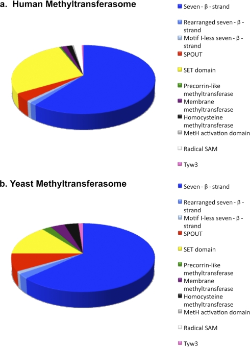
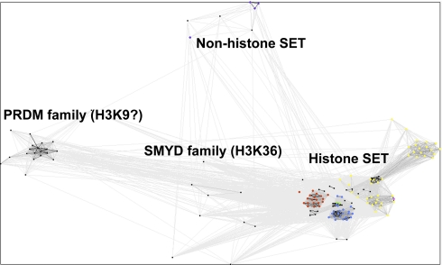
We present a comprehensive analysis of the human methyltransferasome. Primary sequences, predicted secondary structures, and solved crystal structures of known methyltransferases were analyzed by hidden Markov models, Fisher-based statistical matrices, and fold recognition prediction-based threading algorithms to create a model, or profile, of each methyltransferase superfamily. These profiles were used to scan the human proteome database and detect novel methyltransferases. 208 proteins in the human genome are now identified as known or putative methyltransferases, including 38 proteins that were not annotated previously. To date, 30% of these proteins have been linked to disease states. Possible substrates of methylation for all of the SET domain and SPOUT methyltransferases as well as 100 of the 131 seven-β-strand methyltransferases were surmised from sequence similarity clusters based on alignments of the substrate-specific domains.


Figures

Fig. 1.
Fig. 1.
Bioinformatics approaches for searching proteome for methyltransferase family members. Detecting putative methyltransferases requires multiple bioinformatics approaches depending on the existing information for each reference group of superfamily members. An overall scheme of these methods is depicted here; a fuller description is given in the text. Previous bioinformatics searches for novel methyltransferases have used the MEME and MAST programs (4).
Fig. 2.
Fig. 2.
Composition of methyltransferasome. Proteins in the human (a) and yeast (b) methyltransferasomes are sorted into their superfamilies. The composition of the human methyltransferasome was taken from Table I; the yeast methyltransferasome was obtained from Ref. . The radical SAM proteins identified previously in yeast (6) were not included here because they are more closely related to radical SAM non-methyltransferases than to radical SAM methyltransferases.
Fig. 3.
Fig. 3.
Homologs between yeast and human seven-β-strand methyltransferases. The Venn diagram represents the numbers of proteins that have homologs in both human and yeast or are exclusive to each organism (see supplemental Tables IV and V). Proteins that are exclusive either to human or to yeasts are described by function. Although there is poor sequence similarity, yeast Mtq1 is the functional homolog of human HemK.
Fig. 4.
Fig. 4.
Sequence similarity cluster of SPOUT methyltransferases. All of the known and putative SPOUT methyltransferases were analyzed by CLANS with reference proteins to infer substrate specificity. All human SPOUT methyltransferases are in black and identified with arrows. Reference proteins in the NEP1 subfamily are in red, and those in the TrmH subfamily are in yellow. Yeast proteins YGR283C and YMR310C are in dark and light blue, respectively, and guanine-N1-9-methyltransferase reference proteins are in pink. BLAST correlations are shown with gray lines; lighter shades of gray represent BLAST correlations closer to an E-value of 1, whereas darker shades of gray represent BLAST correlations that are closer to an E-value of 0.
Fig. 5.
Fig. 5.
Sequence similarity clusters of SET domain methyltransferases. All of the known and putative SET domain methyltransferases were analyzed by CLANS with reference proteins to infer substrate specificity. All human SET domain methyltransferases are in black. Reference proteins methylating only H3K4 are in red; those methylating H3K9 are in yellow; those methylating H3K36 are in blue; those methylating both H3K36 and H4K20 are in green; those methylating H3K9, H3K27, and H4K20 are in pink; and those methylating Rubisco are in purple. BLAST correlations are shown with gray lines; lighter shades of gray represent BLAST correlations closer to an E-value of 1, whereas darker shades of gray represent BLAST correlations that are closer to an E-value of 0.

Similar articles

Cited by

References

    1. Cheng X., Blumenthal R. M. (eds) (1999) S-Adenosylmethionine-dependent Methyltransferases: Structures and Functions, World Scientific, Singapore
    1. Martin J. L., McMillan F. M. (2002) SAM (dependent) I AM: the S-adenosylmethionine-dependent methyltransferase fold. Curr. Opin. Struct. Biol. 12, 783–793 - PubMed
    1. Schubert H. L., Blumenthal R. M., Cheng X. (2003) Many paths to methyltransfer: a chronicle of convergence. Trends Biochem. Sci. 28, 329–335 - PMC - PubMed
    1. Katz J. E., Dlakić M., Clarke S. (2003) Automated identification of putative methyltransferases from genomic open reading frames. Mol. Cell. Proteomics 2, 525–540 - PubMed
    1. Petrossian T. C., Clarke S. G. (2009) Multiple Motif Scanning to identify methyltransferases from the yeast proteome. Mol. Cell. Proteomics 8, 1516–1526 - PMC - PubMed

Publication types

LinkOut - more resources