. 2011 Jan 6;117(1):352-61.
doi: 10.1182/blood-2010-06-291245. Epub 2010 Oct 7.

IL-6 in human cytomegalovirus secretome promotes angiogenesis and survival of endothelial cells through the stimulation of survivin

Affiliations

IL-6 in human cytomegalovirus secretome promotes angiogenesis and survival of endothelial cells through the stimulation of survivin

Sara Botto et al. Blood. .

Abstract

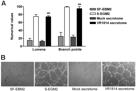
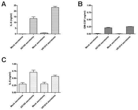
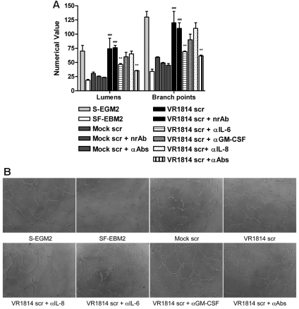
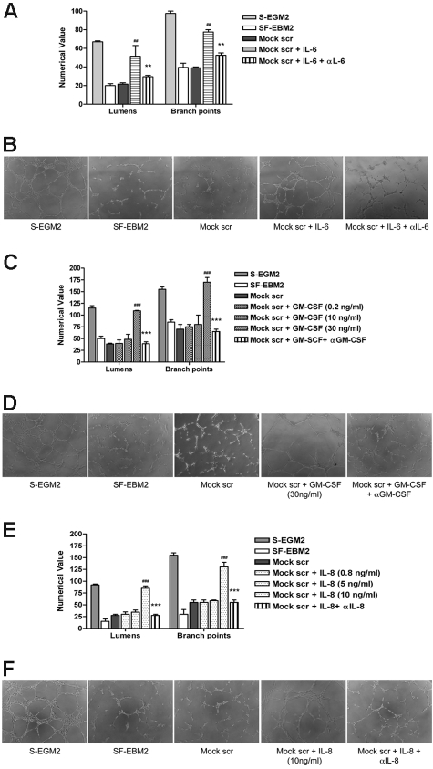
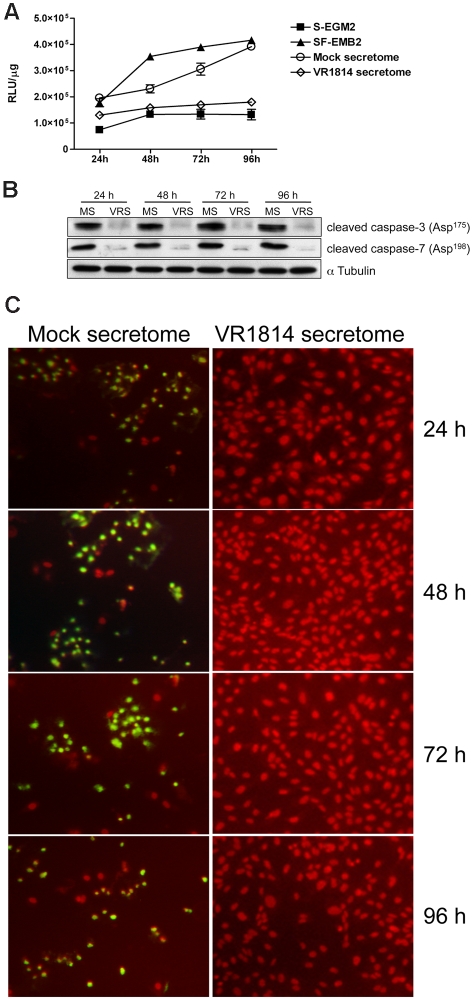
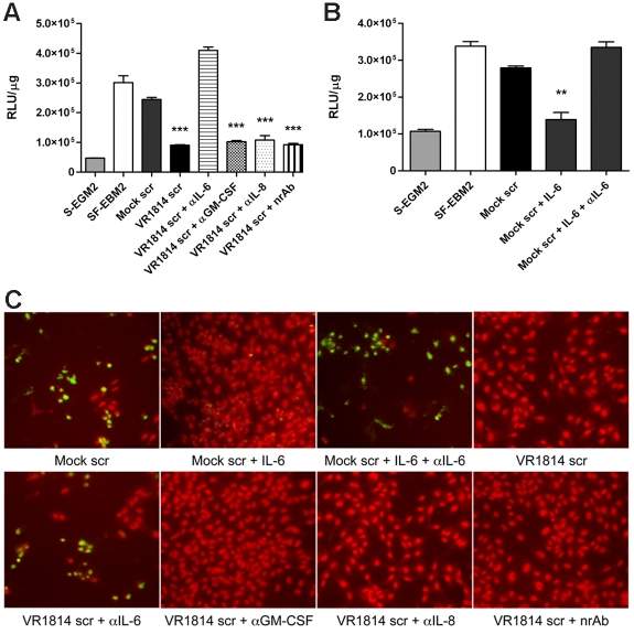
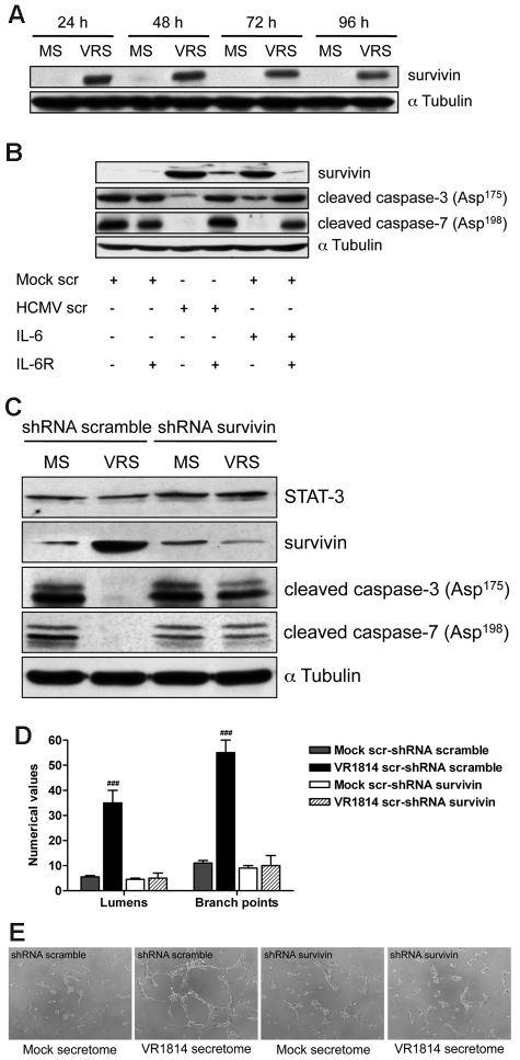
Human cytomegalovirus (HCMV) is linked to the acceleration of vascular diseases such as atherosclerosis and transplant vasculopathy. One of the hallmarks of these diseases is angiogenesis (AG) and neovessel formation. Endothelial cells (ECs) are an integral part of AG and are sites of HCMV persistence. AG requires multiple synchronous processes that include EC proliferation, migration, and vessel stabilization. Virus-free supernatant (secretome) from HCMV-infected ECs induces AG. To identify factor(s) involved in this process, we performed a human cytokine array. Several cytokines were significantly induced in the HCMV secretomes including interleukin-6 (IL-6), granulocyte macrophage colony-stimulating factor, and IL-8/CXCL8. Using in vitro AG assays, neutralization of IL-6 significantly reduced neovessel formation. Addition of the HCMV secretome to preformed vessels extended neovessel survival, but this effect was blocked by neutralization of IL-6. In these cells, IL-6 prevented apoptosis by blocking caspase-3 and -7 activation through the induction of survivin. Neutralization of IL-6 receptor on ECs abolished the ability of HCMV secretome to increase survivin expression and activated effector caspases. Moreover, survivin shRNA expression induced rapid regression of tubule capillary networks in ECs stimulated with HCMV secretome and activated effector caspases. These observations may explain how CMV accelerates vascular disease despite limited infection in tissues.


Figures

Figure 1
Figure 1
Supernatants from HCMV-infected ECs induce AG. (A) Quantitative measures of AG consisted of the number of lumens and branch points. The results are expressed as mean ± SD, ##P < .01 versus mock. (B) Representative examples of each culture condition are shown as a low-power image (magnification, ×10).
Figure 2
Figure 2
HCMV induces secretion of the angiogenic factors IL-6, GM-CSF, and IL-8. (A) ELISA quantification of IL-6 levels in 3 different supernatants of fibroblasts infected with HCMV AD169 and HUVECs infected with HCMV VR1814. (B) ELISA quantification of GM-CSF levels in 3 different supernatants of fibroblasts infected with HCMV AD169 and HUVECs infected with HCMV VR1814. (C) ELISA quantification of IL-8/CXCL8 levels in 3 different supernatants of fibroblasts infected with HCMV AD169 and HUVECs infected with HCMV VR1814.
Figure 3
Figure 3
Neutralization of IL-6 decreases AG and vessel stabilization at 1 week. (A) Quantification of Matrigel tube formation assay after 24 hours in the presence of mock secretome (Mock scr), mock secretome with a nonrelated antibody (Mock scr + nrAb), mock secretome with anti–IL-6, anti–GM-CSF, and anti–IL-8/CXCL8 (Mock scr + αAbs), HCMV VR1814 secretome (VR1814 scr), HCMV VR1814 secretome with a nonrelated antibody (VR1814 scr + nrAb), HCMV VR1814 secretome with anti–IL-6 antibody (VR1814 scr + αIL-6), HCMV VR1814 secretome with anti–GM-CSF antibody (VR1814 scr + αGM-CSF), HCMV VR1814 secretome with anti–IL-8/CXCL8 antibody (VR1814 scr + αIL-8), and HCMV VR1814 secretome with anti–IL-6, anti–GM-CSF, and anti–IL-8/CXCL8 antibodies (VR1814 scr + αAbs). The results are expressed as mean ± SD, ###P < .001 versus mock, **P < .01 versus HCMV VR1814 secretome. (B) Representative examples of each culture conditions are shown as a low-power image (magnification, ×10). (C) Tubule survival is shown after 1 week on Matrigel in the presence of mock secretome, HCMV VR1814 secretome, HCMV VR1814 secretome with anti–IL-6 antibody (VR1814 scr + αIL-6) at low-power image (magnification, ×10), and at high-power image (magnification, ×20).
Figure 4
Figure 4
Exogenous IL-6, GM-CSF, and IL-8 promote HUVEC tube formation. (A) Quantification of Matrigel tube formation assay after 24 hours in the presence of human recombinant IL-6 (10 ng/mL). The results are expressed as mean ± SD, ##P < .01 versus mock, **P < .01 versus mock + IL-6. (B) Representative examples of each culture condition are shown as a low-power image (magnification, ×10). (C) Quantification of Matrigel tube formation assay after 24 hours in the presence of increasing concentrations of GM-CSF. The results are expressed as mean ± SD, ###P < .001 versus mock, ***P < .001 versus mock + GM-CSF (30 ng/mL). (D) Representative examples of each culture condition are shown as a low-power image (magnification, ×10). (E) Quantification of Matrigel tube formation assay after 24 hours in the presence of increasing concentrations of IL-8/CXCL8. The results are expressed as mean ± SD, ##P < .01 versus mock, **P < .01 versus mock + IL-8/CXCL8. (F) Representative examples of each culture condition are shown as a low-power image (magnification, ×10).
Figure 5
Figure 5
HCMV secretomes decrease caspase-3 and -7 activity. HUVECs were starved overnight and then stimulated with mock- or HCMV-secretomes during a time course analysis. (A) Cytosolic extracts from HUVECs incubated with controls, mock- and HCMV VR1814-secretome for 24, 48, 72, and 96 hours were prepared in hypotonic extraction buffer. An equal volume of a proluminescent substrate (DEVD), and cytosolic proteins were added to a white-walled 96-well plate and incubated at room temperature for 1 hour. The luminescence of each sample run in triplicate was measured in a plate-reading luminometer. The graphic shows the mean ± SD of 3 independent experiments. (B) Cell lysates from HUVECs incubated with mock-secretome (MS) and HCMV VR1814 (VRS) were subjected to SDS-PAGE followed by Western blotting to evaluate the expression of activated/cleaved caspase-3 and -7. αTubulin served as an internal control. (C) HUVECs were stimulated as above, and nuclear DNA fragmentation as a sign of apoptosis was determined over the time using a TUNEL assay. Fragmented DNA is indicated by green fluorescence. Nuclei were stained with propidium iodide (red fluorescence).
Figure 6
Figure 6
Neutralization of IL-6 activity in the HCMV secretomes induces apoptosis. HUVECs were starved overnight and then stimulated with mock-secretome, HCMV-secretome, and neutralized HCMV-secretomes for 48 hours. (A) Cytosolic extracts from HUVECs incubated with controls, mock-secretome, HCMV VR1814-secretome, and HCMV VR1814-secretome neutralized with anti–IL-6, anti–GM-CSF, anti–IL-8/CXCL8, or nonrelated antibody were prepared in hypotonic extraction buffer. An equal volume of a proluminescent substrate (DEVD) and cytosolic proteins were added to a white-walled 96-well plate and incubated at room temperature for 1 hour. The luminescence of each sample run in triplicate was measured in a plate-reading luminometer. The results are expressed as mean ± SD of 3 independent experiments, ***P < .001 versus mock. (B) HUVECs were starved overnight and then stimulated with mock-secretome, mock-secretome with IL-6 (10 ng/mL), and mock-secretome with IL-6 (10 ng/mL) + anti–IL-6 Ab (2 μg/mL) for 48 hours. Cytosolic extracts were prepared as above and the luminescence of each sample run in triplicate was measured in a plate-reading luminometer. The results are expressed as mean ± SD of 3 independent experiments, **P < .01 versus mock. (C) TUNEL assay performed in the same experimental conditions as above. Fragmented DNA is indicated by green fluorescence. Nuclei were stained with propidium iodide (red fluorescence).
Figure 7
Figure 7
HCMV secretome stimulates survivin expression through IL-6 receptor blocking apoptosis and promoting AG. (A) Cell lysates from HUVECs incubated with mock-secretome (MS) and HCMV VR1814 (VRS) for 24, 48, 72, and 96 hours were subjected to SDS-PAGE followed by Western blotting to evaluate the expression of survivin. αTubulin served as an internal control. (B) HUVECs were treated with anti–IL-6R antibody and stimulated with the secretome for 48 hours. Expression of survivin, cleaved caspase-3, and caspase-7 was determined by Western blotting. αTubulin immunodetected with mAb served as an internal control. (C) HUVECs were infected with recombinant retroviruses expressing either a survivin-directed shRNA or a nonspecific control (scramble) shRNA followed by stimulation with the HCMV secretome. Expression of STAT-3, survivin, cleaved caspase-3, and caspase-7 was determined by Western blotting. αTubulin served as an internal control. (D) Quantification of Matrigel tube formation assay after 24 hours of HUVECs in the same experimental condition as above. The results are expressed as mean ± SD, ###P < .01 versus mock. (B) Representative examples of each culture condition are shown as a low-power image (magnification, ×10).

Similar articles

Cited by

References

    1. Hosenpud JD, Shipley GD, Wagner CR. Cardiac allograft vasculopathy: current concepts, recent developments, and future directions. J Heart Lung Transplant. 1992;11(1 Pt 1):9–23. - PubMed
    1. Zhou YF, Leon MB, Waclawiw MW, et al. Association between prior cytomegalovirus infection and the risk of restenosis after coronary atherectomy. N Engl J Med. 1996;335(9):624–630. - PubMed
    1. Speir E, Modali R, Huang ES, et al. Potential role of human cytomegalovirus and p53 interaction in coronary restenosis. Science. 1994;265(5170):391–394. - PubMed
    1. Streblow DN, Soderberg-Naucler C, Vieira J, et al. The human cytomegalovirus chemokine receptor US28 mediates vascular smooth muscle cell migration. Cell. 1999;99(5):511–520. - PubMed
    1. Melnick JL, Adam E, DeBakey ME. The link between CMV and atherosclerosis. Infect Med. 1998;15:479–486.

Publication types

MeSH terms