

Importance of nonenteric protozoan infections in immunocompromised people
- PMID: 20930074
- PMCID: PMC2952979
- DOI: 10.1128/CMR.00001-10
Importance of nonenteric protozoan infections in immunocompromised people
Abstract
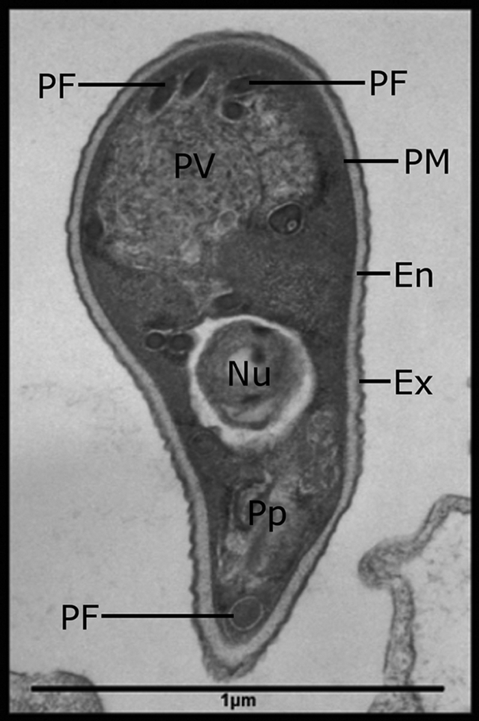
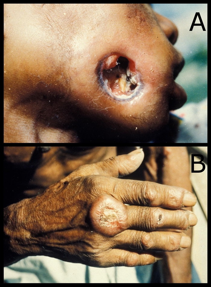
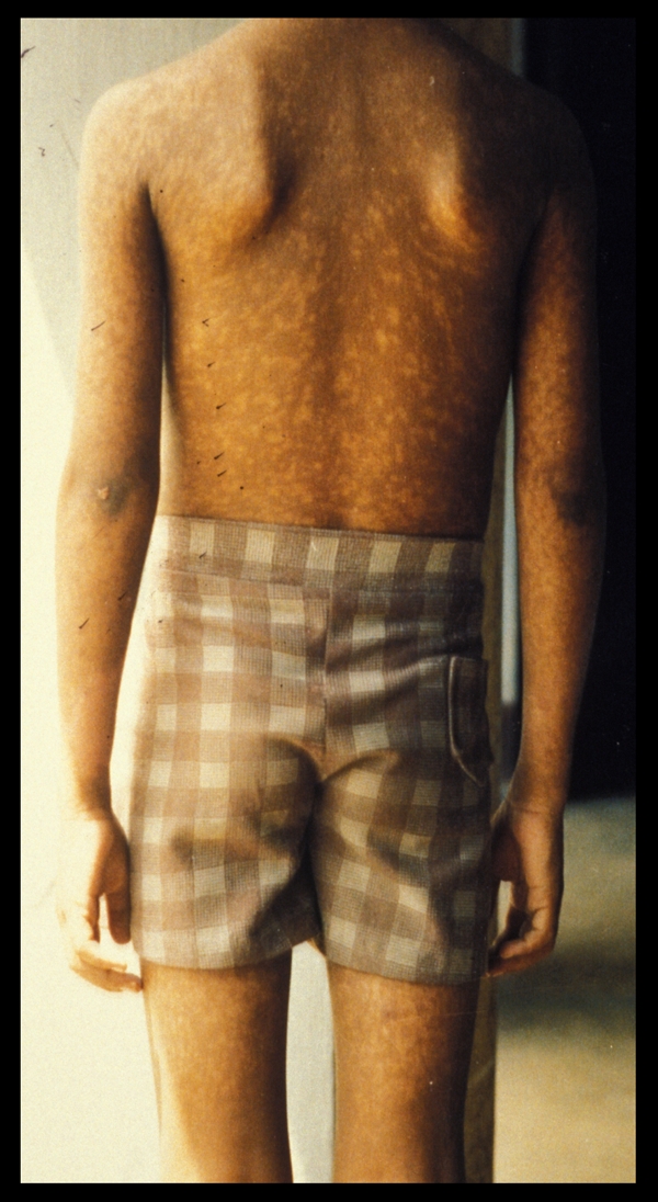
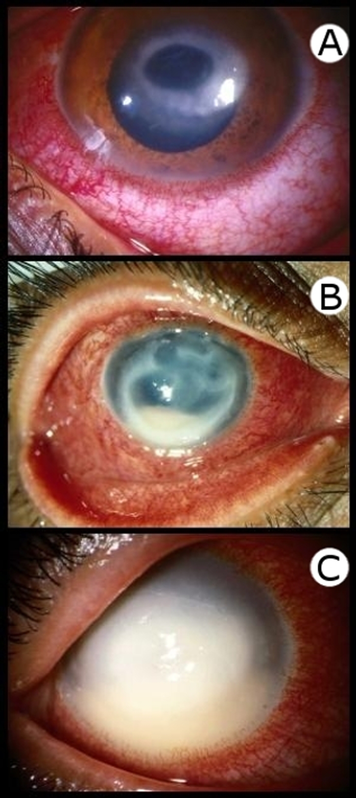
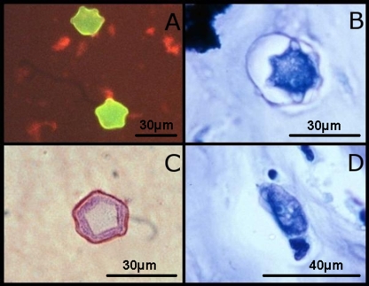
There are many neglected nonenteric protozoa able to cause serious morbidity and mortality in humans, particularly in the developing world. Diseases caused by certain protozoa are often more severe in the presence of HIV. While information regarding neglected tropical diseases caused by trypanosomatids and Plasmodium is abundant, these protozoa are often not a first consideration in Western countries where they are not endemic. As such, diagnostics may not be available in these regions. Due to global travel and immigration, this has become an increasing problem. Inversely, in certain parts of the world (particularly sub-Saharan Africa), the HIV problem is so severe that diseases like microsporidiosis and toxoplasmosis are common. In Western countries, due to the availability of highly active antiretroviral therapy (HAART), these diseases are infrequently encountered. While free-living amoebae are rarely encountered in a clinical setting, when infections do occur, they are often fatal. Rapid diagnosis and treatment are essential to the survival of patients infected with these organisms. This paper reviews information on the diagnosis and treatment of nonenteric protozoal diseases in immunocompromised people, with a focus on patients infected with HIV. The nonenteric microsporidia, some trypanosomatids, Toxoplasma spp., Neospora spp., some free-living amoebae, Plasmodium spp., and Babesia spp. are discussed.
Figures

References
-
- Abad-Franch, F., A. Paucar, C. Carpio, C. A. Cuba, H. M. Aguilar, and M. A. Miles. 2001. Biogeography of Triatominae (Hemiptera: Reduviidae) in Ecuador: implications for the design of control strategies. Mem. Inst. Oswaldo Cruz 96:611-620. - PubMed
-
- Albrecht, H., H. J. Stellbrink, S. Fenske, H. Schafer, and H. Greten. 1994. Successful treatment of Toxoplasma gondii myocarditis in an AIDS patient. Eur. J. Clin. Microbiol. Infect. Dis. 13:500-504. - PubMed
-
- Aleixo, A. L., E. I. Benchimol, E. de Souza Neves, C. S. Silva, L. C. Coura, and M. R. Amendoeira. 2009. Frequency of lesions suggestive of ocular toxoplasmosis among a rural population in the State of Rio de Janeiro. Rev. Soc. Bras. Med. Trop. 42:165-169. (In Portugese.) - PubMed
-
- Alfonzo, M., D. Blanc, C. Troadec, M. Huerre, M. Eliaszewicz, G. Gonzalez, Y. Koyanagi, and D. Scott-Algara. 2002. Temporary restoration of immune response against Toxoplasma gondii in HIV-infected individuals after HAART, as studied in the hu-PBMC-SCID mouse model. Clin. Exp. Immunol. 129:411-419. - PMC - PubMed
Publication types
MeSH terms
LinkOut - more resources
Full Text Sources
Research Materials
Miscellaneous
