Review
. 2010 Oct;23(4):858-83.
doi: 10.1128/CMR.00007-10.

Penetration of drugs through the blood-cerebrospinal fluid/blood-brain barrier for treatment of central nervous system infections

Affiliations
Review

Penetration of drugs through the blood-cerebrospinal fluid/blood-brain barrier for treatment of central nervous system infections

Roland Nau et al. Clin Microbiol Rev. 2010 Oct.

Abstract

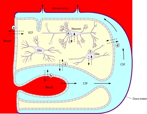
The entry of anti-infectives into the central nervous system (CNS) depends on the compartment studied, molecular size, electric charge, lipophilicity, plasma protein binding, affinity to active transport systems at the blood-brain/blood-cerebrospinal fluid (CSF) barrier, and host factors such as meningeal inflammation and CSF flow. Since concentrations in microdialysates and abscesses are not frequently available for humans, this review focuses on drug CSF concentrations. The ideal compound to treat CNS infections is of small molecular size, is moderately lipophilic, has a low level of plasma protein binding, has a volume of distribution of around 1 liter/kg, and is not a strong ligand of an efflux pump at the blood-brain or blood-CSF barrier. When several equally active compounds are available, a drug which comes close to these physicochemical and pharmacokinetic properties should be preferred. Several anti-infectives (e.g., isoniazid, pyrazinamide, linezolid, metronidazole, fluconazole, and some fluoroquinolones) reach a CSF-to-serum ratio of the areas under the curves close to 1.0 and, therefore, are extremely valuable for the treatment of CNS infections. In many cases, however, pharmacokinetics have to be balanced against in vitro activity. Direct injection of drugs, which do not readily penetrate into the CNS, into the ventricular or lumbar CSF is indicated when other effective therapeutic options are unavailable.


Figures

FIG. 1.
FIG. 1.
Intracranial fluid compartments. Continuous arrows represent the direction of the CSF flow. Interrupted arrows indicate where a diffusion of water or solutes can occur between brain capillaries, CSF, and nervous tissue: (a) across the blood-brain barrier; (b) across the epithelium of the choroid plexus; (c) across the ependyma; (d) across the pia-glial membranes at the surface of the brain and spinal cord; (e and f) across the cell membranes of neurons and glial cells. The thick line represents the dura mater and arachnoidea surrounding the system. (Reproduced from reference with permission of Churchill Livingstone.)
FIG. 2.
FIG. 2.
Drug exchange between blood and CSF. (A) Intravenous injection of a small hydrophilic compound moderately entering the CNS. Shown are semilogarithmic concentration-versus-time curves of fosfomycin in serum (open circles) and ventricular CSF (filled circles) after the intravenous application of 10 g. The AUCCSF-to-AUCS ratio was 0.138. The concentrations in CSF lagged behind those in serum, and the elimination half-life in CSF was considerably longer than that in serum, suggesting that the drug was eliminated mainly by bulk flow. (Reproduced from reference with permission from Urban & Vogel, Munich, Germany.) (B) Double-logarithmic hysteresis loops illustrating the CSF-to-serum concentration ratios of the relatively hydrophilic fluoroquinolone ciprofloxacin over 24 h after intravenous infusion of 200 mg. The concentration ratio increased with the interval between infusion and CSF and serum sampling in three patients (open squares, closed squares, and closed triangles). Please note that this ratio can attain almost any value depending on the interval between infusion and measurement. (Reproduced from reference with permission of Oxford University Press.) (C) Intravenous injection of a moderately lipophilic fluoroquinolone with a relatively low molecular mass readily crossing the blood-CSF barrier. Shown are concentration-versus-time curves in serum and ventricular CSF of ofloxacin and its metabolites after the intravenous application of 400 mg. Please note that after the initial distribution phase, serum (red filled circles) and CSF (red open circles) ofloxacin concentrations almost ran in parallel, indicating that CSF bulk flow was negligible compared to diffusion across the blood-CSF barrier. The overall penetration of ofloxacin into the CSF was greater than that of its more hydrophilic metabolites ofloxacin-N-oxide (serum, blue filled circles; CSF, blue open circles) and N-desmethyl-ofloxacin (serum, green filled circles; CSF, green open circles) (AUCCSF0-24 h/AUCS0-24 h of 0.62 ± 0.09 versus 0.14 ± 0.10 and 0.37 ± 0.35; P < 0.05). (Reproduced from reference .) (D) Intraventricular injection of a large hydrophilic compound with low entry into the CNS when administered systemically (vancomycin). Hour 0 denotes the time point immediately before the intraventricular (re)injection of 10 mg vancomycin. Columns represent means ± standard deviations (SD) of data from three patients. Please note that therapeutic CSF levels are encountered over 24 h. (Reproduced from reference with permission of the University of Chicago Press.)
FIG. 2.
FIG. 2.
Drug exchange between blood and CSF. (A) Intravenous injection of a small hydrophilic compound moderately entering the CNS. Shown are semilogarithmic concentration-versus-time curves of fosfomycin in serum (open circles) and ventricular CSF (filled circles) after the intravenous application of 10 g. The AUCCSF-to-AUCS ratio was 0.138. The concentrations in CSF lagged behind those in serum, and the elimination half-life in CSF was considerably longer than that in serum, suggesting that the drug was eliminated mainly by bulk flow. (Reproduced from reference with permission from Urban & Vogel, Munich, Germany.) (B) Double-logarithmic hysteresis loops illustrating the CSF-to-serum concentration ratios of the relatively hydrophilic fluoroquinolone ciprofloxacin over 24 h after intravenous infusion of 200 mg. The concentration ratio increased with the interval between infusion and CSF and serum sampling in three patients (open squares, closed squares, and closed triangles). Please note that this ratio can attain almost any value depending on the interval between infusion and measurement. (Reproduced from reference with permission of Oxford University Press.) (C) Intravenous injection of a moderately lipophilic fluoroquinolone with a relatively low molecular mass readily crossing the blood-CSF barrier. Shown are concentration-versus-time curves in serum and ventricular CSF of ofloxacin and its metabolites after the intravenous application of 400 mg. Please note that after the initial distribution phase, serum (red filled circles) and CSF (red open circles) ofloxacin concentrations almost ran in parallel, indicating that CSF bulk flow was negligible compared to diffusion across the blood-CSF barrier. The overall penetration of ofloxacin into the CSF was greater than that of its more hydrophilic metabolites ofloxacin-N-oxide (serum, blue filled circles; CSF, blue open circles) and N-desmethyl-ofloxacin (serum, green filled circles; CSF, green open circles) (AUCCSF0-24 h/AUCS0-24 h of 0.62 ± 0.09 versus 0.14 ± 0.10 and 0.37 ± 0.35; P < 0.05). (Reproduced from reference .) (D) Intraventricular injection of a large hydrophilic compound with low entry into the CNS when administered systemically (vancomycin). Hour 0 denotes the time point immediately before the intraventricular (re)injection of 10 mg vancomycin. Columns represent means ± standard deviations (SD) of data from three patients. Please note that therapeutic CSF levels are encountered over 24 h. (Reproduced from reference with permission of the University of Chicago Press.)
FIG. 3.
FIG. 3.
Effects of inhibitors of drug efflux pumps on the penetration of antibiotics into the central nervous system. (A) Concentration of radioactively labeled benzylpenicillin (BP) in the cisterna magna (CM) and the cisterna basalis (CB) after its application into the CM in control and probenecid-pretreated dogs. Concentrations of benzylpenicillin (means ± standard errors of the means [SEM]; n = 4) in the CM and CB over time (minutes), expressed as the percentage of the concentration in the CM at 5 min after drug application. Inhibition of the efflux pump by probenecid increases the CSF concentrations by a factor of 3 to 15. (Reproduced from reference with permission of Elsevier B.V.) (B) Increased drug concentrations in P-glycoprotein knockout mice (149). The percent increases (concentration in knockout mice divided by the concentration in wild-type mice) in brain (dark blue bars) and plasma (light blue bars) for different drugs investigated by Schinkel and coworkers are shown. Contrarily to brain levels, plasma concentrations were not changed substantially, indicating a key role for P-gp at the blood-brain/blood-CSF barrier. (Reproduced from reference with permission of the American Society for Pharmacology and Experimental Therapeutics.)

References

    1. Agwuh, K. N., and A. MacGowan. 2006. Pharmacokinetics and pharmacodynamics of the tetracyclines including glycylcyclines. J. Antimicrob. Chemother. 58:256-265. - PubMed
    1. Albanese, J., M. Leone, B. Bruguerolle, M. L. Ayem, B. Lacarelle, and C. Martin. 2000. Cerebrospinal fluid penetration and pharmacokinetics of vancomycin administered by continuous infusion to mechanically ventilated patients in an intensive care unit. Antimicrob. Agents Chemother. 44:1356-1358. - PMC - PubMed
    1. Albert, F., G. B. Bishop-Freudling, and H. Vergin. 1984. Diffusion of tetroxoprim and sulfadiazine in the cerebrospinal fluid of neurosurgery patients. Fortschr. Med. 102:1064-1066. (In German.) - PubMed
    1. Alffenaar, J. W., P. M. de Vries, G. J. Luijckx, D. van Soolingen, T. S. van der Werf, and R. van Altena. 2008. Plasma and cerebrospinal fluid pharmacokinetics of moxifloxacin in a patient with tuberculous meningitis. Antimicrob. Agents Chemother. 52:2293-2295. - PMC - PubMed
    1. Alffenaar, J. W., R. van Altena, H. J. Bokkerink, G. J. Luijckx, D. van Soolingen, R. E. Aarnoutse, and T. S. van der Werf. 2009. Pharmacokinetics of moxifloxacin in cerebrospinal fluid and plasma in patients with tuberculous meningitis. Clin. Infect. Dis. 49:1080-1082. - PubMed

Publication types

MeSH terms

Substances

LinkOut - more resources