Review
. 2010 Oct;23(4):884-928.
doi: 10.1128/CMR.00019-10.

Melanized fungi in human disease

Affiliations
Review

Melanized fungi in human disease

Sanjay G Revankar et al. Clin Microbiol Rev. 2010 Oct.

Erratum in

  • Clin Microbiol Rev. 2012 Oct;25(4):720

Abstract

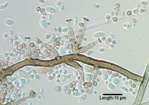
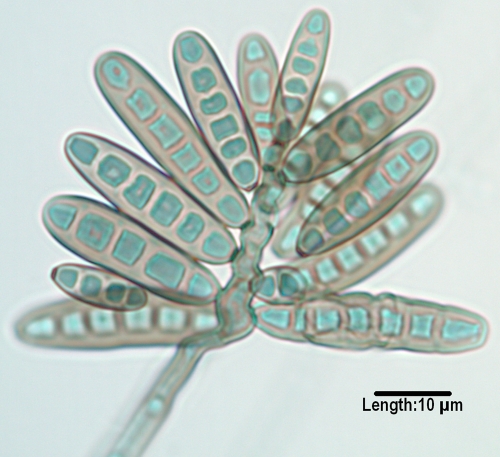
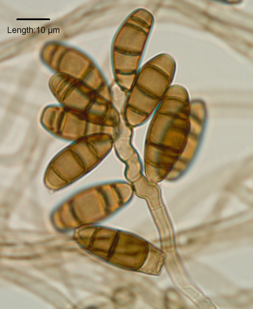
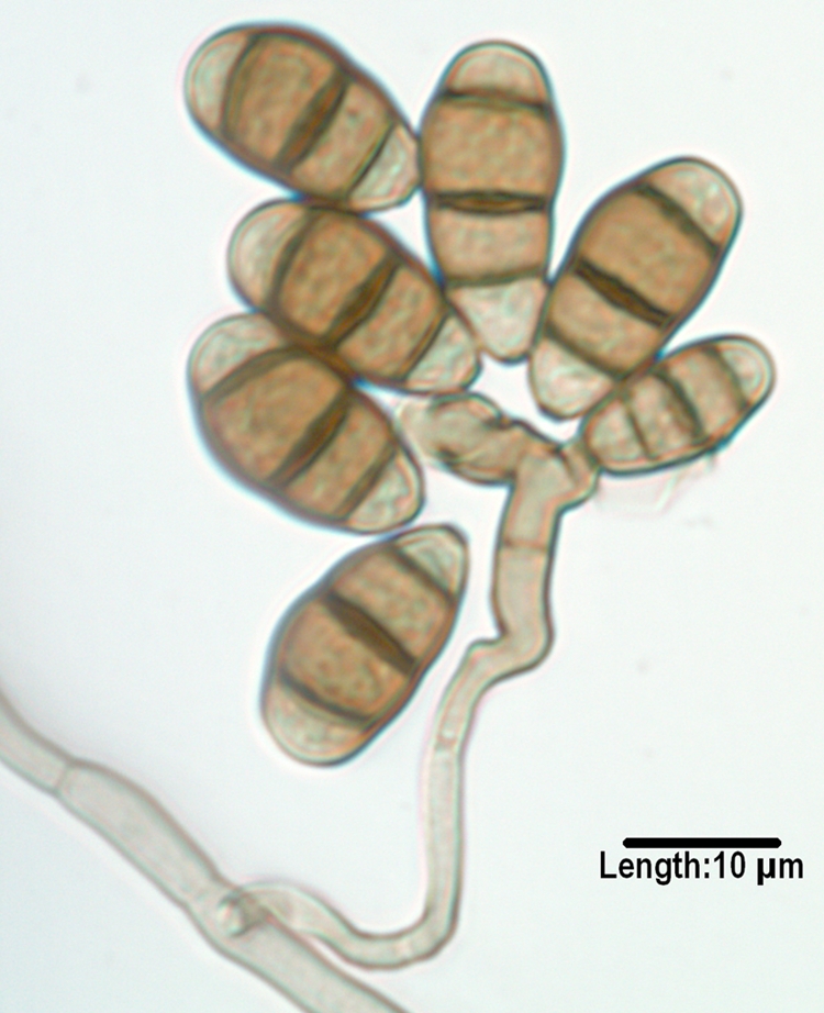
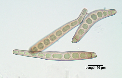
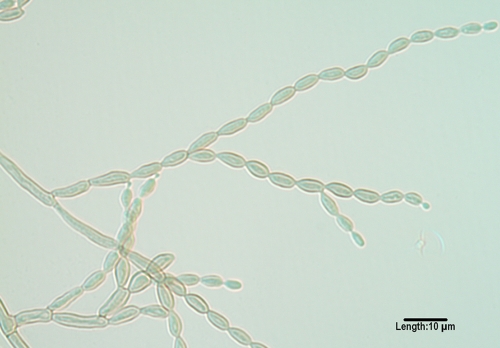
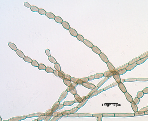
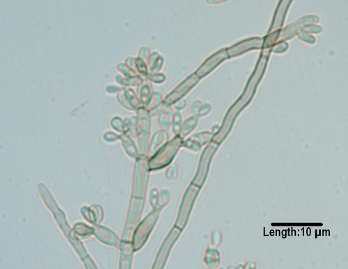
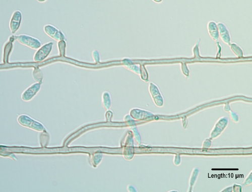
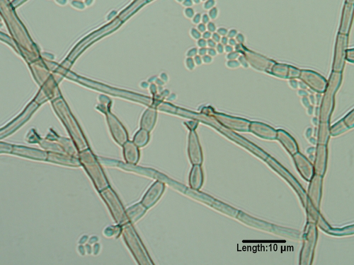
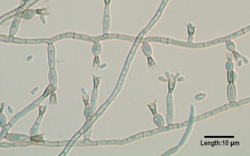
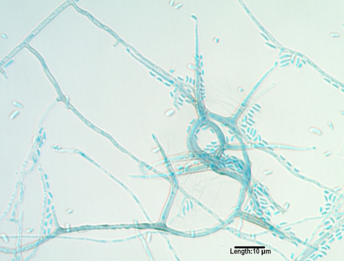
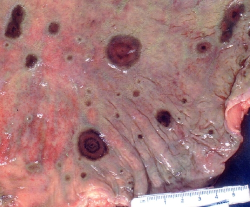
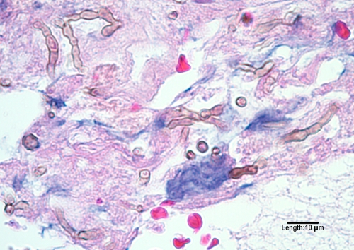
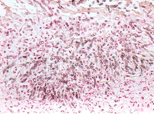
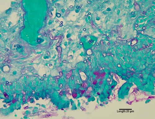
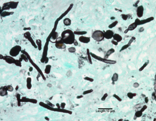
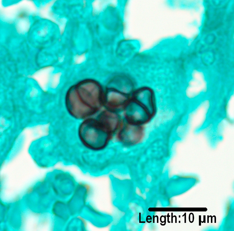
Melanized or dematiaceous fungi are associated with a wide variety of infectious syndromes, including chromoblastomycosis, mycetoma, and phaeohyphomycosis. [corrected]. Many are soil organisms and are generally distributed worldwide, though certain species appear to have restricted geographic ranges. Though they are uncommon causes of disease, melanized fungi have been increasingly recognized as important pathogens, with most reports occurring in the past 20 years. The spectrum of diseases with which they are associated has also broadened and includes allergic disease, superficial and deep local infections, pneumonia, brain abscess, and disseminated infection. For some infections in immunocompetent individuals, such as allergic fungal sinusitis and brain abscess, they are among the most common etiologic fungi. Melanin is a likely virulence factor for these fungi. Diagnosis relies on careful microscopic and pathological examination, as well as clinical assessment of the patient, as these fungi are often considered contaminants. Therapy varies depending upon the clinical syndrome. Local infection may be cured with excision alone, while systemic disease is often refractory to therapy. Triazoles such as voriconazole, posaconazole, and itraconazole have the most consistent in vitro activity. Further studies are needed to better understand the pathogenesis and optimal treatment of these uncommon infections.


Figures

FIG. 1.
FIG. 1.
Conidiogenous cells of Pleurostomophora richardsiae, demonstrating prominent flaring collarettes as well as the two types of conidia (oval and globose) produced by this species. (Unless otherwise noted, in this and subsequent figures light microscopy photomicrographs of conidiogenous cells and/or conidia were taken from slide culture preparations grown on potato flakes agar for 7 days at 25°C.)
FIG. 2.
FIG. 2.
Conidia of Bipolaris hawaiiensis, demonstrating mostly five distosepta and six cells being borne from a geniculate conidiophore/conidiogeous cell.
FIG. 3.
FIG. 3.
Conidia of Curvularia inaequalis with mostly five septa and six cells borne from a geniculate conidiogenous cells.
FIG. 4.
FIG. 4.
Conidia of Curvularia lunata var. aeria borne from a geniculate conidiogenous cell. Note that the middle cell is slightly enlarged, and septa are eusepta (true septa continuous with the outer wall).
FIG. 5.
FIG. 5.
Multidistoseptate conidia of Exserohilum longirostratum, demonstrating a prominent basal septum (true septum) and a protruding hilum.
FIG. 6.
FIG. 6.
Long, nonfragile chains of conidia as seen in Cladophialophora bantiana.
FIG. 7.
FIG. 7.
Long, nonfragile chains of conidia produced by a less common species of Cladophialophora, C. emmonsii. Note that conidiophores and prominent hila (attachment scars) are absent.
FIG. 8.
FIG. 8.
Conidial formation in Fonsecaea monophora. Conidia are formed from swollen denticles which give rise to secondary and tertiary conidia in chains of up to four conidia. The same type of conidiogenesis occurs in F. pedrosoi.
FIG. 9.
FIG. 9.
Two-celled, clavate (club-shaped) conidia of Ochroconis gallopava borne on long, thin denticles.
FIG. 10.
FIG. 10.
Dark, funnel-shaped collarettes at the tips of the conidiogenous cells (phialides) in Phialophora verrucosa. Also note the oval-shaped conidia.
FIG. 11.
FIG. 11.
Deep, dark, vase-shaped collarettes in Phialophora americana.
FIG. 12.
FIG. 12.
Melanized hyphae, demonstrating warts (bottom), long robust phialides, and allantoid (curved) conidia of Phaeoacremonium parasiticum.
FIG. 13.
FIG. 13.
A GMS-stained cross section of a multilocular pycnidium of a Microsphaeropsis species produced on carnation leaf agar after 5 weeks of incubation at 25°C.
FIG. 14.
FIG. 14.
Perithecium of Microascus trigonosporus formed on potato flake agar after 3 weeks of incubation at 25°C. Note ascospores being released from the ostiole in the neck of the perithecium.
FIG. 15.
FIG. 15.
Heart-shaped ascospores of Microascus cirrosus produced on potato flake agar after 3 weeks of incubation at 25°C.
FIG. 16.
FIG. 16.
Triangular ascospores of Microascus trigonosporus produced on potato flake agar after 3 weeks of incubation at 25°C.
FIG. 17.
FIG. 17.
Bipolaris spicifera colonies in stomach mucosa of patient with disseminated disease (autopsy). (Reproduced from reference [original Fig. 15-6A] with kind permission of Springer Science and Business Media.)
FIG. 18.
FIG. 18.
H&E stain of melanized, moniliform hyphal elements of Cladophialophora bantiana from a brain abscess.
FIG. 19.
FIG. 19.
Bipolaris spicifera in lung tissue (Fontana-Masson stain; magnification ×100). (Reproduced from reference [original Fig. 15-13B] with kind permission of Springer Science and Business Media.)
FIG. 20.
FIG. 20.
PAS stain of Ochrocladosporium elatum, formerly Cladosporium elatum, from sinus tissue.
FIG. 21.
FIG. 21.
GMS stain of Rhinocladiella mackenziei from a brain abscess. Note the many moniform hyphal elements often seen with melanized fungi.
FIG. 22.
FIG. 22.
GMS stain of sclerotic bodies produced by Fonsecaea pedrosoi.

References

    1. Abbott, S. P., L. Sigler, R. McAleer, D. A. McGough, M. G. Rinaldi, and G. Mizell. 1995. Fatal cerebral mycoses caused by the ascomycete Chaetomium strumarium. J. Clin. Microbiol. 33:2692-2698. - PMC - PubMed
    1. Abliz, P., K. Fukushima, K. Takizawa, and K. Nishimura. 2004. Identification of pathogenic dematiaceous fungi and related taxa based on large subunit ribosomal DNA D1/D2 domain sequence analysis. FEMS Immunol. Med. Microbiol. 40:41-49. - PubMed
    1. Adam, R. D., M. L. Paquin, E. A. Petersen, M. A. Saubolle, M. G. Rinaldi, J. G. Corcoran, J. N. Galgiani, and R. E. Sobonya. 1986. Phaeohyphomycosis caused by the fungal genera Bipolaris and Exserohilum. A report of 9 cases and review of the literature. Medicine 65:203-217. - PubMed
    1. Adeyemi, O. A., O. Lie, R. Bernstein, N. Gottardi-Littell, K. Muro, D. Patil, and G. A. Noskin. 2007. Woman with multiple brain abscesses. Clin. Infect. Dis. 45:1351-1352:1397-1399. - PubMed
    1. Agarwal, R. 2009. Allergic bronchopulmonary aspergillosis. Chest 135:805-826. - PubMed