

Large artery calcification on dialysis patients is located in the intima and related to atherosclerosis
- PMID: 20930091
- PMCID: PMC3052220
- DOI: 10.2215/CJN.04290510
Large artery calcification on dialysis patients is located in the intima and related to atherosclerosis
Abstract
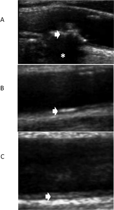
Background and objectives: Vascular calcification (VC) has a significant effect in cardiovascular diseases on dialysis patients. However, VC is assessed with x-ray-based techniques, which do not inform about calcium localization (intima, media, atherosclerosis-related). The aim of this work is to study VC and its related factors using arterial ultrasound to report the exact location of calcium.
Design, setting, participants, & measurements: This was an observational, cross-sectional, case-control study that included 232 patients in dialysis and 208 age- and sex-matched controls with normal kidney function. Demographic data and laboratory values were collated. Carotid, femoral, and brachial ultrasounds were performed to assess VC and atherosclerosis burden using a standardized protocol.
Results: Cardiovascular risk factors were predominantly found in controls, although the burden of atherosclerosis was higher in the dialysis group. VC was significantly more prevalent in the group of patients on dialysis than control subjects, and in both groups the most prevalent pattern of VC was linear calcification located in the intima of the artery wall. Age and undergoing dialysis (with or without previous cardiovascular diseases) were positively and significantly associated with linear calcification. Conversely, the absence of atherosclerosis and low levels of C-reactive protein and phosphorus significantly impeded the development of linear calcification.
Conclusions: VC in large, conduit arteries is more prevalent in patients on dialysis than controls and is predominantly located in a linear fashion in the intima of the arteries.
Figures
References
-
- Reslerova M, Moe SM: Vascular calcification in dialysis patients: Pathogenesis and consequences. Am J Kidney Dis 41(Suppl 1): S96–S99, 2003 - PubMed
-
- Okuno S, Ishimura E, Kitatani K, Fujino Y, Kohno K, Maeno Y, Maekawa K, Yamakawa T, Imanishi Y, Inaba M, Nishizawa Y: Presence of abdominal aortic calcification is significantly associated with all-cause and cardiovascular mortality in maintenance hemodialysis patients. Am J Kidney Dis 49: 417–425, 2007 - PubMed
-
- London GM, Guérin AP, Marchais SJ, Métivier F, Pannier B, Adda H: Arterial media calcification in end-stage renal disease: Impact on all-cause and cardiovascular mortality. Nephrol Dial Transplant 18: 1731–1740, 2003 - PubMed
-
- Adragão T, Pires A, Birne R, Curto JD, Lucas C, Gonçalves M, Negrão AP: A plain x-ray vascular calcification score is associated with arterial stiffness and mortality in dialysis patients. Nephrol Dial Transplant 24: 997–1002, 2009 - PubMed
-
- Kauppila LI, Polak JF, Cupples LA, Hannan MT, Kiel DP, Wilson PW: New indices to classify location, severity and progression of calcific lesions in the abdominal aorta: A 25-year follow-up study. Atherosclerosis 132: 245–250, 1997 - PubMed
Publication types
MeSH terms
Substances
LinkOut - more resources
Full Text Sources
Medical
Research Materials
