Review
. 2010 Oct 15;123(Pt 20):3425-34.
doi: 10.1242/jcs.067611.

Tubulin depolymerization may be an ancient biological motor

Affiliations
Review

Tubulin depolymerization may be an ancient biological motor

J Richard McIntosh et al. J Cell Sci. .

Abstract

The motions of mitotic chromosomes are complex and show considerable variety across species. A wealth of evidence supports the idea that microtubule-dependent motor enzymes contribute to this variation and are important both for spindle formation and for the accurate completion of chromosome segregation. Motors that walk towards the spindle pole are, however, dispensable for at least some poleward movements of chromosomes in yeasts, suggesting that depolymerizing spindle microtubules can generate mitotic forces in vivo. Tubulin protofilaments that flare outward in association with microtubule shortening may be the origin of such forces, because they can move objects that are appropriately attached to a microtubule wall. For example, some kinetochore-associated proteins can couple experimental objects, such as microspheres, to shortening microtubules in vitro, moving them over many micrometers. Here, we review recent evidence about such phenomena, highlighting the force-generation mechanisms and different coupling strategies. We also consider bending filaments of the tubulin-like protein FtsZ, which form rings girding bacteria at their sites of cytokinesis. Mechanical similarities between these force-generation systems suggest a deep phylogenetic relationship between tubulin depolymerization in eukaryotic mitosis and FtsZ-mediated ring contraction in bacteria.


Figures

Fig. 1.
Fig. 1.
Fission yeast chromosomes move poleward in the absence of pole-directed motor enzymes. (A,B) Cells were deprived of MTs by growing a cold-sensitive mutant of β-tubulin at a restrictive temperature (18°C) for 6 hours; this caused the kinetochores to lose their pole-proximal association (Grishchuk and McIntosh, 2006). Upon reversion to permissive conditions (32°C), MTs grew from the centrosomes and encountered the now-dispersed chromosomes. Subsequent chromosome motions were monitored using fluorescence microscopy. (A) Three S. pombe cells from a 3- to 10-minute sample showing stages in chromosome attachment to the spindle. White arrows suggest an inferred progression. Chromosomes, purple; centrosomes, green; kinetochores, red. (B) Fluorescence imaging of a living cell in which both poles (green arrowheads) and one kinetochore (red arrowheads) were tagged with GFP. Imaging was done over a period of 10 minutes. SPB, spindle pole body. (C) Four-dimensional microscopy allowed quantification of the rate of chromosome-to-pole motion in different mutant strains of motor proteins. The maximal rate of chromosome motion was unchanged in those strains that lack the kinesin 14 family members Pkl1 and Klp2 as well as the dynein heavy chain (Dhc1) (pkl1Δklp2Δdhc1Δ; green), revealing that these motors are not at the root of poleward chromosome motion. (D,E) Although the absence of minus-end-directed motors had a relatively minor impact on mitotic progression in wild-type cells (D), the motors contributed to the accuracy and expediency of the poleward motion and bi-orientation when cells were challenged by chromosome scatter (E). Adapted from Grishchuk and McIntosh with permission (Grishchuk and McIntosh, 2006). Chromosomes, purple; centrosomes, green; kinetochores, red (for panels A,D,E).
Fig. 2.
Fig. 2.
Tubulin depolymerization exerts force on inert objects coupled tightly to a MT wall. (A,B) Experimental setup to measure the depolymerization force. MTs initiated from coverslip-bound nucleators grow with their plus ends distal. A microsphere coated with streptavidin binds to biotinylated MT segments (blue) and can be captured in a laser trap (red cone) as well as monitored with a quadrant photodiode (not shown). Although Brownian movement renders the position of the bead variable (see photodiode data in B), MT depolymerization pulls the bead briefly, moving it away from the center of the trap until the bead-associated tubulin is fully depolymerized. The bead then relaxes to the center of the trap. (C) Relative size of bead (0.25 μm radius in this drawing) and the MT. The lateral mode of the attachment of the bead, the large distance between the MT attachment site (where the force is exerted) and the center of the bead (where the force is measured with a laser trap) cause the measured force to be less than that developed by the bending PFs. (D) Consistent with the interpretation given in C, beads of smaller diameter report a larger force (compare black horizontal bars). Yellow vertical bars indicate the range of force predicted theoretically. Adapted from Grishchuk et al. (Grishchuk et al., 2008a).
Fig. 3.
Fig. 3.
The Dam1 complex forms rings and smaller oligomers that can serve as processive couplers to MT depolymerization. (A) EM images (right panels) of a negatively stained MT that is either attached to an immobilizing film, followed by incubation without (top) and with soluble Dam1 (middle) or that is not attached and incubated together with soluble Dam1 (bottom). (Left panels) Schematic of the same experimental setup. (B,C) Fluorescent studies reveal that all Dam1 oligomers (but not stacks of multiple rings) exhibit some diffusion and move with a shortening MT end; but their rates of diffusion (B) and tracking shortening MTs (C) decrease with increasing oligomer size (error bars in B are ± s.e.m.; curve in C is a hyperbolic fit to the data and vertical bar at each point shows the 95% confidence level). (D) Dam1-coated beads follow the shortening MT ends by two distinct mechanisms, depending on the presence of soluble Dam1. (E) Large oligomers allow a shortening MT to push on a bead with a force that is approximately five times greater than that seen with a biotin–avidin linkage. Shown are unprocessed quadrant photodiode records from a Dam1-coated bead in the presence of soluble Dam1 and a streptavidin-coated bead (inset). Adapted from Grishchuk et al. (Grishchuk et al., 2008b).
Fig. 4.
Fig. 4.
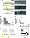
Bead tracking assays to identify kinetochore–MT couplers. (A) Microbeads coated with the Ndc80 complex attach to tethered MTs (left panel) and follow their ends as they shorten (right panel); time-lapse images of a moving bead, adapted from McIntosh et al. with permission (McIntosh et al., 2008). (B) Fluorescent Ndc80 molecules diffuse on a MT wall in a low-ionic-strength buffer, as illustrated with a kymograph. Reproduced with permission from Powers et al. (Powers et al., 2009). (C) Comparison of the coupling properties of different kinetochore proteins (for more details see supplementary material Table S1). By the two criteria of percent processive beads (red bars) and mean interaction time (green bars and ± s.e.m.), the Dam1 complex provides the most efficient bead transport, followed by its presumptive ortholog, the Ska1-containing complex.
Fig. 5.
Fig. 5.
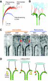
Fibrillar structures between kinetochores and spindle MTs, and their possible role in force-transducing coupling. (A) The average shape of PFs from kinetochore-associated MTs (KMTs) is intermediate between those of polymerizing and depolymerizing MTs. (B) This shape may be caused by fibrillar attachments between the bending PFs and chromosomes, which serve as a load; drawing based on electron tomography (fibrils, red; MTs, green; chromatin, blue). (C) Fibrils connect these intermediate protofilaments with chromatin in averaged images of kinetochore-associated MTs from cultured mammalian cells (red arrows). The more-bent PFs, called ‘Ram's horns’ lack these connections in averaged images. Adapted from McIntosh et al. with permission (McIntosh et al., 2008). (D) Consecutive stages in this hypothetical coupling; non-diffusing fibrils pull on the attached cargo because of power-strokes from bending PFs.
Fig. 6.
Fig. 6.
Organization and function of FstZ filaments in eubacteria. (A) Fusion of FtsZ protein with GFP forms a band at the mid-region of a bacterial cell. This band decreases in diameter as the cell cleaves (image kindly provided by J. Lutkenhaus, University of Kansas, Kansas City, KS). (B) Purified FtsZ–YFP chimera, engineered to include a membrane-binding domain. This protein induces indentations (arrows) in tubular liposomes in vitro, demonstrating its sufficiency for bending membranes. (C) The same liposome imaged before and after a 6-minute interval, scale bar 5 μm. Adapted from Osawa et al. with permission (Osawa et al., 2008). (D) Slices cut from cryo-electron tomograms of cleaving Caulobacter cells that are overexpressing FtsZ. Straight and curved segments of FtsZ filaments are visible in these 6.7-nm slices. Abrupt kinks are sometimes seen (black arrows) as well as direct connections of straight filaments to the membrane (white arrowheads); scale bar 50 nm. Adapted from Li et al. with permission (Li et al., 2007). (E) Summary of curvature-dependent cellular functions of bending tubulin-like proteins (blue) and their attaching links (red) to a cargo (black).

Similar articles

Cited by

References

    1. Asbury C. L., Gestaut D. R., Powers A. F., Franck A. D., Davis T. N. (2006). The Dam1 kinetochore complex harnesses microtubule dynamics to produce force and movement. Proc. Natl. Acad. Sci. USA 103, 9873-9878 - PMC - PubMed
    1. Brinkley B. R., Stubblefield E. (1966). The fine structure of the kinetochore of a mammalian cell in vitro. Chromosoma 19, 28-43 - PubMed
    1. Cassimeris L., Inoue S., Salmon E. D. (1988). Microtubule dynamics in the chromosomal spindle fiber: analysis by fluorescence and high-resolution polarization microscopy. Cell Motil. Cytoskeleton 10, 185-196 - PubMed
    1. Cheeseman I. M., Brew C., Wolyniak M., Desai A., Anderson S., Muster N., Yates J. R., Huffaker T. C., Drubin D. G., Barnes G. (2001). Implication of a novel multiprotein Dam1p complex in outer kinetochore function. J. Cell Biol. 155, 1137-1145 - PMC - PubMed
    1. Cheeseman I. M., Chappie J. S., Wilson-Kubalek E. M., Desai A. (2006). The conserved KMN network constitutes the core microtubule-binding site of the kinetochore. Cell 127, 983-997 - PubMed

Publication types