

Ets1 blocks terminal differentiation of keratinocytes and induces expression of matrix metalloproteases and innate immune mediators
- PMID: 20930145
- PMCID: PMC2959112
- DOI: 10.1242/jcs.062240
Ets1 blocks terminal differentiation of keratinocytes and induces expression of matrix metalloproteases and innate immune mediators
Abstract
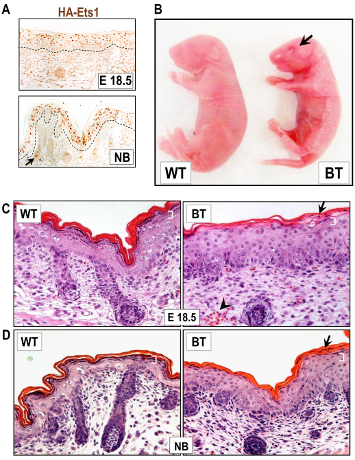
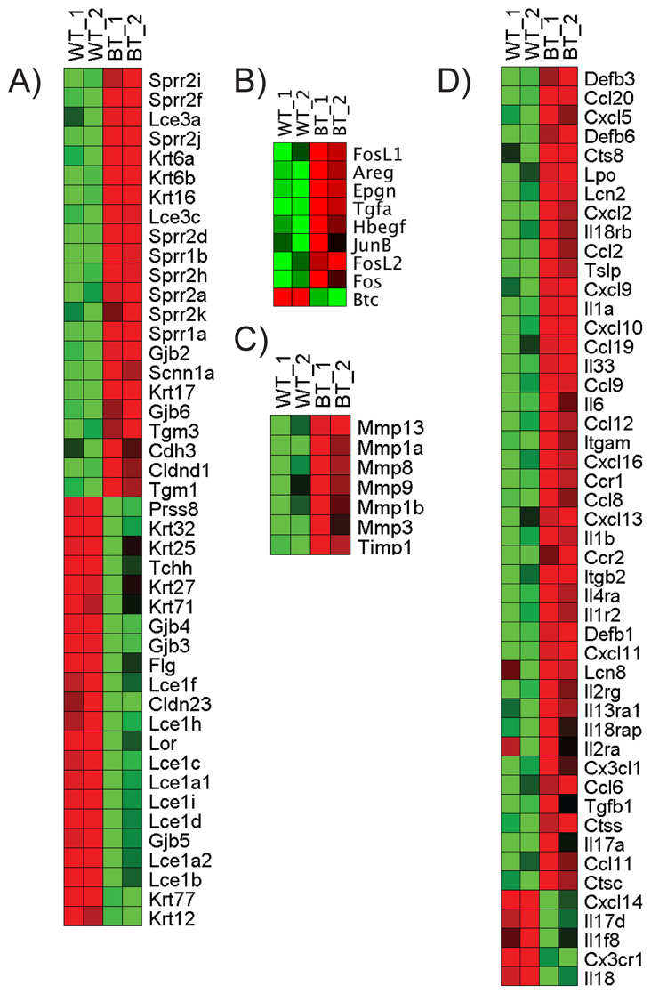
The transcription factor Ets1 is normally expressed in the proliferative layer of stratified epithelium, but expression of Ets1 is significantly upregulated in squamous cell carcinomas. How elevated levels of Ets1 impact tumor initiation and progression is not well understood. To determine the biological consequences of overexpression of Ets1, we developed a transgenic mouse model that allows induction of Ets1 expression in keratinocytes of stratified epithelium in a regulatable fashion. Induction of Ets1 during embryonic development results in a dramatic alteration in epidermal structure and function by suppressing the expression of multiple stratum corneum constituents, while at the same time inducing expression of EGF ligands, AP1 transcription factors and matrix metalloproteases. Interestingly, expression of certain immune-related genes, including defensins, chemokines and cytokines was increased as well, suggesting a possible role for immune dysregulation in the promotion of squamous dysplasia. Experiments using cultured mouse keratinocytes indicate that Ets1 can induce expression of some of these mediators in a cell-intrinsic fashion. Collectively, our data reveal that elevated expression of Ets1 has a much broader array of pro-tumorigenic effects on epithelial cells than previously appreciated.
Figures


Similar articles
-
Unique domain functions of p63 isotypes that differentially regulate distinct aspects of epidermal homeostasis.Carcinogenesis. 2006 Jan;27(1):53-63. doi: 10.1093/carcin/bgi200. Epub 2005 Aug 4. Carcinogenesis. 2006. PMID: 16081516
-
Mesenchymal stem cells differentiate into keratinocytes and express epidermal kallikreins: Towards an in vitro model of human epidermis.J Cell Biochem. 2019 Aug;120(8):13141-13155. doi: 10.1002/jcb.28589. Epub 2019 Mar 19. J Cell Biochem. 2019. PMID: 30891818
-
Role of the Notch ligand Delta1 in embryonic and adult mouse epidermis.J Invest Dermatol. 2008 Apr;128(4):825-32. doi: 10.1038/sj.jid.5701113. Epub 2007 Oct 25. J Invest Dermatol. 2008. PMID: 17960184
-
An update on the roles of transcription factor Ets1 in autoimmune diseases.WIREs Mech Dis. 2023 Nov-Dec;15(6):e1627. doi: 10.1002/wsbm.1627. Epub 2023 Aug 10. WIREs Mech Dis. 2023. PMID: 37565573 Free PMC article. Review.
-
Innate immunity and the regulation and mobilization of keratinocyte stem cells: are the old players playing a new game?Exp Dermatol. 2012 Sep;21(9):660-4. doi: 10.1111/j.1600-0625.2012.01566.x. Exp Dermatol. 2012. PMID: 22897573 Free PMC article. Review.
Cited by
-
AIRE is induced in oral squamous cell carcinoma and promotes cancer gene expression.PLoS One. 2020 Feb 3;15(2):e0222689. doi: 10.1371/journal.pone.0222689. eCollection 2020. PLoS One. 2020. PMID: 32012175 Free PMC article.
-
Analysis of zebrafish periderm enhancers facilitates identification of a regulatory variant near human KRT8/18.Elife. 2020 Feb 7;9:e51325. doi: 10.7554/eLife.51325. Elife. 2020. PMID: 32031521 Free PMC article.
-
Differentiation-associated genes regulated by c-Jun and decreased in the progression of esophageal squamous cell carcinoma.PLoS One. 2014 May 5;9(5):e96610. doi: 10.1371/journal.pone.0096610. eCollection 2014. PLoS One. 2014. PMID: 24796531 Free PMC article.
-
Identification of functional non-coding variants associated with orofacial cleft.Nat Commun. 2025 Jul 16;16(1):6545. doi: 10.1038/s41467-025-61734-w. Nat Commun. 2025. PMID: 40670354 Free PMC article.
-
The Role of p16INK4a Pathway in Human Epidermal Stem Cell Self-Renewal, Aging and Cancer.Int J Mol Sci. 2017 Jul 22;18(7):1591. doi: 10.3390/ijms18071591. Int J Mol Sci. 2017. PMID: 28737694 Free PMC article. Review.
References
-
- Akiyama M., Smith L. T., Yoneda K., Holbrook K. A., Hohl D., Shimizu H. (1999). Periderm cells form cornified cell envelope in their regression process during human epidermal development. J. Invest. Dermatol. 112, 903-909 - PubMed
-
- Ala-aho R., Kahari V. M. (2005). Collagenases in cancer. Biochimie 87, 273-286 - PubMed
-
- Aoki T., Kataoka H., Nishimura M., Ishibashi R., Morishita R., Miyamoto S. (2010). Ets-1 promotes the progression of cerebral aneurysm by inducing the expression of MCP-1 in vascular smooth muscle cells. Gene Ther. Epub ahead of print - PubMed
-
- Arlette J. P., Trotter M. J. (2004). Squamous cell carcinoma in situ of the skin: history, presentation, biology and treatment. Austr. J. Dermatol. 45, 1-9; quiz 10 - PubMed
Publication types
MeSH terms
Substances
Grants and funding
LinkOut - more resources
Full Text Sources
Molecular Biology Databases
Miscellaneous
