. 2010 Nov;11(11):854-60.
doi: 10.1038/embor.2010.151. Epub 2010 Oct 8.

Trichoplein/mitostatin regulates endoplasmic reticulum-mitochondria juxtaposition

Affiliations

Trichoplein/mitostatin regulates endoplasmic reticulum-mitochondria juxtaposition

Cristina Cerqua et al. EMBO Rep. 2010 Nov.

Abstract

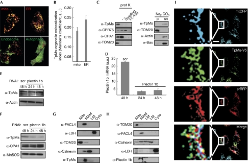
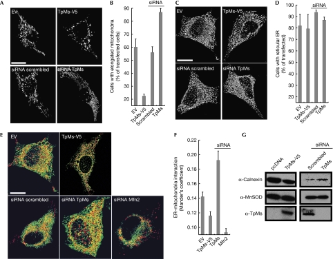
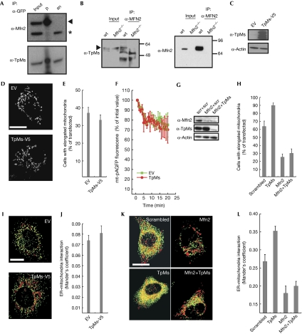
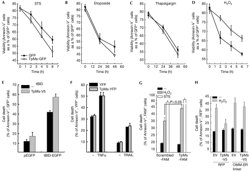
Trichoplein/mitostatin (TpMs) is a keratin-binding protein that partly colocalizes with mitochondria and is often downregulated in epithelial cancers, but its function remains unclear. In this study, we report that TpMs regulates the tethering between mitochondria and endoplasmic reticulum (ER) in a Mitofusin 2 (Mfn2)-dependent manner. Subcellular fractionation and immunostaining show that TpMs is present at the interface between mitochondria and ER. The expression of TpMs leads to mitochondrial fragmentation and loosens tethering with ER, whereas its silencing has opposite effects. Functionally, the reduced tethering by TpMs inhibits apoptosis by Ca(2+)-dependent stimuli that require ER-mitochondria juxtaposition. Biochemical and genetic evidence support a model in which TpMs requires Mfn2 to modulate mitochondrial shape and tethering. Thus, TpMs is a new regulator of mitochondria-ER juxtaposition.


Conflict of interest statement

The authors declare that they have no conflict of interest.

Figures

Figure 1
Figure 1
Trichoplein/mitostatin is enriched at the endoplasmic reticulum–mitochondria interface. (A) Upper panels: representative confocal images of HeLa cells co-transfected with TpMs–GFP (green) and mtRFP (mito, red) or erRFP (ER, red). Lower panels: confocal images of HeLa cells co-transfected with TpMs–V5 and empty vector (left) or YFP-LC3 (autophago, right), fixed after 24 h and immunostained with TRITC-conjugated anti-V5 (red) and with an FITC-conjugated cM6PR antibody (endosomes, green) in the left panel. Scale bar, 20 μm. (B) Mean±s.e. (n=3) of interaction data from (A). (C) Left: crude HeLa mitochondria (5 mg/ml) were incubated where indicated with proteinase K (100 μg/ml). Mitochondria were in isolation buffer (Frezza et al, 2007) or in 20 mM HEPES (pH 7.4; swelling) or in 0.1% Triton X-100. Proteins (25 μg) separated by SDS–PAGE were immunoblotted with the indicated antibodies. Right: crude HeLa mitochondria were incubated in 0.1 M Na2CO3 (pH 11.3; 30 min; 4°C). After centrifugation, proteins (25 μg) from pellet (p) and supernatant (sn) separated by SDS–PAGE were immunoblotted with the indicated antibodies. (D) Real-time PCR of plectin 1b levels from HeLa cells transfected with the indicated siRNA. (E) Proteins (20 μg) from HeLa cells transfected as indicated were separated by SDS–PAGE and immunoblotted using the indicated antibodies. (F) Mitochondria were isolated at indicated times from 5 × 10⁸ HeLa cells transfected as indicated and proteins (25 μg) were separated by SDS–PAGE and immunoblotted. (G,H) Proteins (40 μg) from Percoll-purified subcellular fractions of mouse liver, were separated by SDS–PAGE and immunoblotted. (I) Representative confocal images of HeLa cells co-transfected with the indicated plasmids. After 24 h cells were fixed and immunostained with FITC-conjugated V5 antibody. Boxed areas are magnified × 9. Scale bar, 20 μm. White in the merge image: overlap of the three channels. Cyto, cytosol; ER, endoplasmic reticulum; ER-RFP, ER-targeted red fluorescent protein; FITC, fluorescein isothiocyanate; GFP, green fluorescent protein; GRP75, glucose regulatory protein 75; LC3, microtuble-associated protein 1 light chain 3; LM, light membranes; MAM, mitochondria-associated membrane; mito, mitochondria; mtRFP, matrix red fluorescent protein; SDS–PAGE, sodium dodecyl sulphate–polyacrylamide gel electrophoresis; scr, scrambled; siRNA, small interfering RNA; TpMs, trichoplein/mitostatin; TRITC, tetramethyl rhodamine iso-thiocyanate; YFP, yellow fluorescent protein.
Figure 2
Figure 2
Trichoplein/mitostatin regulates mitochondrial shape and mitochondria–endoplasmic reticulum juxtaposition. (A) Representative confocal images of HeLa cells co-transfected with mtRFP and the indicated plasmids. Scale bar, 20 μm. (B) Mean±s.e.m. (n=3) of morphometric analysis from (A). (C) Representative three-dimensional reconstructions of ER HeLa cells co-transfected with ER-YFP and the indicated plasmids. Scale bar, 20 μm. (D) Mean±s.e.m. (n=3) of morphometric analysis from (C). (E) Representative three-dimensional reconstructions of ER and mitochondria in HeLa cells co-transfected with mtRFP, ER-YFP and the indicated plasmids. Yellow, organelles are closer than around 270 nm. Scale bar, 20 μm. (F) Mean±s.e. (n=5) of interaction data from (E). (G) A total of 25 μg of proteins from mitochondria isolated after 24 (left) or 48 h (right) from HeLa cells (5 × 10⁸), transfected as indicated, were analysed by SDS–PAGE and immunoblotting. ER, endoplasmic reticulum; ER-RFP, ER-targeted red fluorescent protein; Mfn2, mitofusin 2; MtRFP, matrix red fluorescent protein; SDS–PAGE, sodium dodecyl sulphate–polyacrylamide gel electrophoresis; siRNA, small interfering RNA; TpMs, trichoplein/mitostatin; YFP, yellow fluorescent protein.
Figure 3
Figure 3
Trichoplein/mitostatin physically and functionally interacts with mitofusin 2. (A) Proteins (300 μg) from pre-cleared lysates of HeLa cells transfected with Mfn2–GFP were immunoprecipitated as indicated and analysed by SDS–PAGE and immunoblotting. Input and supernatant are diluted 1:10. (B) Proteins (1 mg) from pre-cleared lysates of wt and Mfn2−/− MEFs were immunoprecipitated as indicated and analysed by SDS–PAGE and immunoblotting. Input and supernatant are diluted 1:10. (C) Proteins (20 μg) from Mfn2−/− MEFs 24 h after transfection with the indicated plasmids were analysed by SDS–PAGE and immunoblotting. (D) Representative confocal images of Mfn2−/− MEFs co-transfected for 24 h with mtRFP and the indicated plasmids. Scale bar, 20 μm. (E) Mean±s.e. (n=3) of morphometric analysis of mitochondrial shape from (D). (F) Mitochondrial fusion assay of Mfn2−/− MEFs co-transfected with mtRFP, mt-pAGFP and the indicated plasmids. Data are mean±s.e. (n=3). (G) HeLa cells were co-transfected with the indicated siRNA and after 48 h cells were lysed and protein (20 μg) was analysed by SDS–PAGE and immunoblotting using the indicated antibodies. (H) Mean±s.e. (n=3) of morphometric analysis of mitochondrial shape from HeLa cells transfected with the indicated siRNA and mtRFP. (I) Three-dimensional reconstructions of ER and mitochondria in Mfn2−/− MEFs co-transfected with mtRFP, ER-YFP and the indicated plasmids. Yellow, organelles are closer than about 270 nm. Scale bar, 20 μm. (J) Mean±s.e. (n=3) of interaction data from (G). (K) Three-dimensional reconstructions of ER and mitochondria in HeLa cells co-transfected with mtRFP, ER-YFP and the indicated siRNA. Yellow indicates that organelles are closer than around 270 nm. Scale bar, 20 μm. (L) Mean±s.e. (n=5) of interaction data from (K). ER, endoplasmic reticulum; ER-RFP, ER-targeted red fluorescent protein; EV, empty vector; GFP, green fluorescent protein; MEFs, mouse embryonic fibroblasts; Mfn2, mitofusin 2; mtRFP, matrix red fluorescent protein; pAGFP, green fluorescent protein; SDS–PAGE, sodium dodecyl sulphate–polyacrylamide gel electrophoresis; siRNA, small interfering RNA; wt, wild type; YFP, yellow fluorescent protein.
Figure 4
Figure 4
Trichoplein/mitostatin protects cells from Ca2+-dependent apoptosis. (A–D) Viability of HeLa cells transfected as indicated and treated for the indicated times with staurosporine (STS, 2 μm, A), etoposide (5 μm, B), thapsigargin (1 μm, C) and H2O2 (1 mM, D). (E) Cell death of HeLa cells 48 h after transfection with the indicated and plasmids. (F) Cell death of HeLa cells transfected as indicated and treated with TNF-α (25 ng/ml plus 10 μg/ml cycloheximide) or TRAIL (50 ng/ml) for 16 h. In (AF), data are represented as mean±s.e. (n=5). (G) Cell death of HeLa cells transfected as indicated and treated after 48 h with H2O2 or STS for 6 h. Data are represented as mean±s.e. (n=3). (H) Cell death of HeLa cells co-transfected as indicated and treated after 24 h with H2O2 for a duration of 4 h. Data are represented as mean±s.e. (n=6). EGFP, enhanced green fluorescent protein; ER, endoplasmic reticulum; EV, empty vector; FAM, carboxyfluorescein; OMM, outer mitochondrial membrane; RFP, red fluorescent protein; tBID, truncated BID; TNF, tumour necrosis factor; TpMs, trichoplein/mitostatin; TRAIL, tumour-necrosis-factor-related apoptosis-inducing ligand; YFP, yellow fluorescent protein.

References

    1. Anesti V, Scorrano L (2006) The relationship between mitochondrial shape and function and the cytoskeleton. Biochim Biophys Acta 1757: 692–699 - PubMed
    1. Cipolat S, de Brito OM, Dal Zilio B, Scorrano L (2004) OPA1 requires mitofusin 1 to promote mitochondrial fusion. Proc Natl Acad Sci USA 101: 15927–15932 - PMC - PubMed
    1. Csordas G, Renken C, Varnai P, Walter L, Weaver D, Buttle KF, Balla T, Mannella CA, Hajnoczky G (2006) Structural and functional features and significance of the physical linkage between ER and mitochondria. J Cell Biol 174: 915–921 - PMC - PubMed
    1. de Brito OM, Scorrano L (2008) Mitofusin 2 tethers endoplasmic reticulum to mitochondria. Nature 456: 605–610 - PubMed
    1. Dimmer KS, Navoni F, Casarin A, Trevisson E, Endele S, Winterpacht A, Salviati L, Scorrano L (2008) LETM1, deleted in Wolf Hirschhorn syndrome is required for normal mitochondrial morphology and cellular viability. Hum Mol Genet 17: 201–214 - PubMed

Publication types

LinkOut - more resources