. 2010 Oct 8:10:538.
doi: 10.1186/1471-2407-10-538.

LATS2 is de-methylated and overexpressed in nasopharyngeal carcinoma and predicts poor prognosis

Affiliations

LATS2 is de-methylated and overexpressed in nasopharyngeal carcinoma and predicts poor prognosis

Yan Zhang et al. BMC Cancer. .

Abstract

Background: LATS2, which encodes a novel serine/threonine kinase, is known to be important in centrosome duplication and in the maintenance of genomic stability. Recently, a potential role for LATS2 in cancer has been reported. In breast cancer and acute lymphoblastic leukemia (ALL), LATS2 mRNA is downregulated and has been suggested to be a tumor suppressor. However, the role of LATS2 in nasopharyngeal carcinoma has not been investigated. In this study, we aimed to investigate the expression pattern of LATS2 and its clinicopathological involvement in nasopharyngeal carcinoma to understand its effect on cell survival.

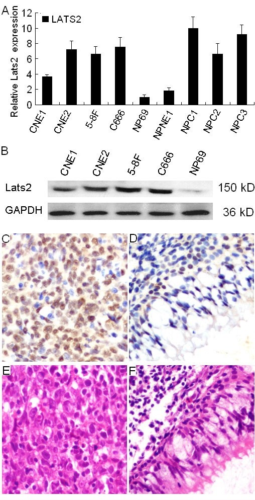
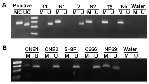
Methods: Using quantitative real time PCR and immunoblotting, the expression of LATS2 was detected in nasopharyngeal carcinoma cell lines and in the immortalized nasopharyngeal epithelial cell line NP69. Using immunohistochemistry, we analyzed LATS2 protein expression in 220 nasopharyngeal carcinoma cases. The association of LATS2 protein expression with the clinicopathological characteristics and the prognosis of nasopharyngeal carcinoma were subsequently assessed. Using methylation specific PCR, we detected the methylation status of the LATS2 promoter. RNA interference was performed by transfecting siRNA to specifically knock down LATS2 expression in 5-8F and CNE2.

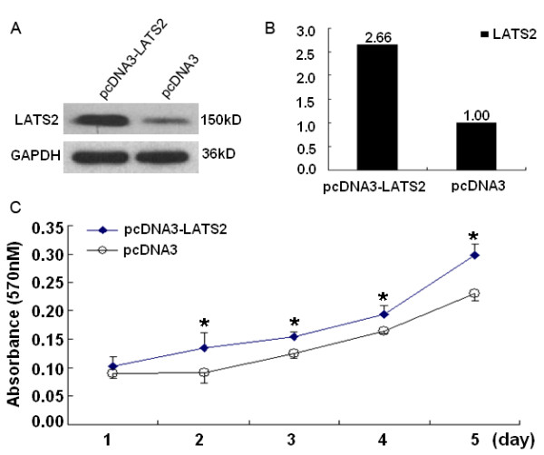
Results: LATS2 protein was detected in 178 of 220 (80.91%) cases of nasopharyngeal carcinoma. LATS2 overexpression was a significant, independent prognosis predictor (P = 0.037) in nasopharyngeal carcinoma patients. Methylation specific PCR revealed that 36.7% (11/30) of nasopharyngeal carcinoma tissues and all of the chronic nasopharyngeal inflammation samples were methylated. Functional studies showed that the suppression of LATS2 expression in nasopharyngeal carcinoma (5-8F and CNE2) cell lines by using specific small interfering (siRNA) resulted in the inhibition of growth, induction of apoptosis and S-phase cell cycle increase. Overexpression of LATS2 in NP69 stimulated cell proliferation.

Conclusions: Our results indicate that LATS2 might play a role in the tumorigenesis of nasopharyngeal carcinoma by promoting the growth of nasopharyngeal carcinoma cells. Transfection with specific siRNA might be feasible for the inhibition of growth, induction of apoptosis and S phase increase in nasopharyngeal carcinoma.


Figures

Figure 1
Figure 1
Expression of LATS2 in NPC cell lines and NPC tumor tissues. A, The expression levels of LATS2 transcripts in the NPC cell lines CNE1, CNE2, 5-8F and C666-1, and in immortalized primary nasopharyngeal epithelial NP69 cells, as well as in three NPC biopsies and one paired normal epithelium were evaluated by quantitative real-time PCR. The NPC cell lines CNE1, CNE2, 5-8F and C666-1, as well as NPC tumors (NPC1, NPC2 and NPC3) show a higher LATS2 mRNA expression level than immortalized primary nasopharyngeal epithelial NP69 cells and normal nasopharynx epithelium (NPNE1). B, The LATS2 protein expression level was detected by western blot in the NPC cell lines CNE1, CNE2, 5-8F, C666-1 cells and in NP69 cells. C, LATS2 (IHC, × 200) staining revealed that overexpression of LATS2 was observed in the nuclear and cytoplasm of carcinoma cells. D, Weak staining of LATS2 was observed in adjacent normal nasopharyngeal epithelial cells. H&E staining of NPC carcinoma cells (E) and adjacent normal nasopharyngeal epithelial cells (F).
Figure 2
Figure 2
Kaplan-Meier survival curves of NPC patients. A, The five-year overall survival rate of 220 NPC patients was 66.18%. B, The five-year overall survival rates were 73.96% and 57.23%, in NPC patients whose tumors showed low levels of LATS2 expression (n = 109) and high levels of LATS2 expression (n = 111), respectively; there was a significant difference in the overall survival rate between the two groups (P = 0.006). C, No significant differences in five-year survival rates were found between low levels of LATS2 expression (n = 29) and high levels of LATS2 expression (n = 25) in NPC patients with early stage disease (stage I - II, P = 0.099). D, The five-year overall survival rates were 70.57% and 57.59% in patients with late stage disease (stage III - IV) whose tumors showed low levels of LATS2 expression (n = 80) and high levels of LATS2 expression (n = 86), respectively; there was a significant difference in the overall survival rate between the two groups (P = 0.032). E, No differences were found in overall survival between low levels of LATS2 expression (n = 28) and high levels of LATS2 expression (n = 30) in NPC patients with differentiated non-keratinising carcinoma (WHO type II, P = 0.125). F, The five-year overall survival rates were 70.91% and 62.23% in patients with undifferentiated carcinoma (WHO type III) whose tumors showed low levels of LATS2 expression (n = 81) and high levels of LATS2 expression (n = 81), respectively; there was a significant difference in the overall survival rate between the two groups (P = 0.040).
Figure 3
Figure 3
Methylation-specific PCR analysis of the LATS2 promoter. Mehtylation of the LATS2 promoter was detected in NPC tumor tissues (A) and NPC cell lines (B). MC, a positive control for methylated alleles. UC, a positive control for unmethylated alleles. M, methylated. U, unmethylated. The methylation-specific products (148 bp) and unmethylation-specific products (130 bp) were separated on 2.5% agarose gels. Marker, 50 bp size marker. T, NPC tumor tissue. N, chronic inflammation of nasopharyngeal samples
Figure 4
Figure 4
LATS2 siRNA1 transfection effectively suppresses LATS2 expression. A, The 5-8F cell line was transfected with three siRNAs (50 nM) to target LATS2 or with control siRNA (50 nM). Cell lysates were generated 72 h post-transfection, followed by immunoblot analysis to determine LATS2 expression; GAPDH was used as the loading control. B, Densitometry analysis revealed that 5-8F cells transfected with LATS2 siRNA1 showed a 78% decrease in LATS2 protein expression compared to cells transfected with control siRNA. C, 5-8F cells were transfected with different concentrations (25 nM, 50 nM, 75 nM and 100 nM) of LATS2 siRNA1 or with control siRNA. Cell lysates were generated at 72 h post-transfection, followed by immunoblot analysis to determine LATS2 expression, GAPDH was used as the loading control. D, Densitometry analysis revealed that 5-8F cells transfected with LATS2 siNRA1 (75 nM and 100 nM) showed a 94% decrease in LATS2 protein expression.
Figure 5
Figure 5
Suppression of LATS2 expression inhibits growth, induces apoptosis and S-phase increase. A, 5-8F and CNE2 cell lines were transfected with LATS2 siRNA1 (75 nM) or control siRNA (75 nM). Cell lysates were generated 72 h post-transfection, followed by immoblot analysis to determine LATS2 expression. GAPDH was used as the loading control. B, Growth curve of 5-8F and CNE2 cells upon LATS2 silencing. Results represent the means ± SD (n = 3).*p < 0.05. C, Flow cytometry analysis of apoptosis by using AnnexinV FITC and PI double staining 72 h after transfection. The percentages of apoptotic 5-8F cells transfected with LATS2 siRNA1 and control siRNA were 42.3% and 18.9%, respectively. In CNE2 cells, the percentages of apoptosis in cells transfected with LATS2 siRNA1 and control siRNA were 34.6% and 17.6%. D. Cell cycle distribution was monitored by flow cytometry. The percentages of cells in S-phase in 5-8F cells transfected with LATS2 siRNA1 and control siRNA were 42.6% and 27.2%, respectively. In CNE2 cells, the percentages of S-phase cells among those transfected with LATS2 siRNA1 and control siRNA were 33.7% and 25.7%, respectively.
Figure 6
Figure 6
Overexpression of LATS2 stimulated cell growth. A, NP69 cells were transfected with pcDNA3-LATS2 or Vector. Cell lysates were generated at 48 h post-transfection, followed by immunoblot analysis to determine LATS2 expression, GAPDH was used as the loading control. B, Densitometry analysis revealed that NP69 cells transfected with pcDNA3-LATS2 showed a 2.66-fold increase in LATS2 protein expression compared to cells transfected with vector. C, MTT assay was used for analysis of the effect of LATS2 on NP69 cell proliferation. Results represent the means ± SD (n = 3).*p < 0.05.

Similar articles

Cited by

References

    1. Chang ET, Adami HO. The enigmatic epidemiology of nasopharyngeal carcinoma. Cancer Epidemiol Biomarkers Prev. 2006;15:1765–77. doi: 10.1158/1055-9965.EPI-06-0353. - DOI - PubMed
    1. Klibi J, Niki T, Riedel A. et al.Blood diffusion and Th1-suppressive effects of galectin-9-containing exosomes released by Epstein-Barr virus-infected nasopharyngeal carcinoma cells. Blood. 2009;113:1957–66. doi: 10.1182/blood-2008-02-142596. - DOI - PubMed
    1. Airoldi M, Gabriele AM, Garzaro M, Induction chemotherapy with cysplatin and epirubicin followed by radiotherapy and concurrent cysplatin in locally advanced nasopharyngeal carcinoma observed in a non-endemic population. Radiother Oncol. 2009. - PubMed
    1. Chen Y, Liu MZ, Liang SB. et al.Preliminary results of a prospective randomized trial comparing concurrent chemoradiotherapy plus adjuvant chemotherapy with radiotherapy alone in patients with locoregionally advanced nasopharyngeal carcinoma in endemic regions of china. Int J Radiat Oncol Biol Phys. 2008;71:1356–64. - PubMed
    1. Farias TP, Dias FL, Lima RA. et al.Prognostic factors and outcome for nasopharyngeal carcinoma. Arch Otolaryngol Head Neck Surg. 2003;129:794–9. doi: 10.1001/archotol.129.7.794. - DOI - PubMed

Publication types

Substances

LinkOut - more resources