. 2010 Oct 8:10:103.
doi: 10.1186/1471-213X-10-103.

PLCγ-activated signalling is essential for TrkB mediated sensory neuron structural plasticity

Affiliations

PLCγ-activated signalling is essential for TrkB mediated sensory neuron structural plasticity

Carla Sciarretta et al. BMC Dev Biol. .

Abstract

Background: The vestibular system provides the primary input of our sense of balance and spatial orientation. Dysfunction of the vestibular system can severely affect a person's quality of life. Therefore, understanding the molecular basis of vestibular neuron survival, maintenance, and innervation of the target sensory epithelia is fundamental.

Results: Here we report that a point mutation at the phospholipase Cγ (PLCγ) docking site in the mouse neurotrophin tyrosine kinase receptor TrkB (Ntrk2) specifically impairs fiber guidance inside the vestibular sensory epithelia, but has limited effects on the survival of vestibular sensory neurons and growth of afferent processes toward the sensory epithelia. We also show that expression of the TRPC3 cation calcium channel, whose activity is known to be required for nerve-growth cone guidance induced by brain-derived neurotrophic factor (BDNF), is altered in these animals. In addition, we find that absence of the PLCγ mediated TrkB signalling interferes with the transformation of bouton type afferent terminals of vestibular dendrites into calyces (the largest synaptic contact of dendrites known in the mammalian nervous system) on type I vestibular hair cells; the latter are normally distributed in these mutants as revealed by an unaltered expression pattern of the potassium channel KCNQ4 in these cells.

Conclusions: These results demonstrate a crucial involvement of the TrkB/PLCγ-mediated intracellular signalling in structural aspects of sensory neuron plasticity.


Figures

Figure 1
Figure 1
Vestibular neurons survive in absence of a functional TrkB/PLCγ site, but nearly all die by disrupting both the TrkB/PLCγ and SHC sites. (A) Total vestibular neuron numbers at P7 in Trkb point mutants (TrkbPLC/PLC; TrkbD/D) compared to controls (TrkbWT/WT; TrkbPLC/+). Shown also is the loss of vestibular neurons in TrkbSHC/SHC point mutants and in Trkb null mice (Trkb-/-) for comparison. (B-D) Representative cresyl violet-stained horizontal sections of the central portion of the vestibular ganglion of control and point mutant (TrkbPLC/PLC; TrkbD/D) mice at P7 as indicated. ***, p < 0.0001 (ANOVA). Scale bar, 50 μm.
Figure 2
Figure 2
Innervations of vestibular and cochlear sensory epithelia are highly compromised in mice lacking both the PLCγ and the SHC docking sites of TrkB. The pattern of afferent (A, D) and efferent (B-C, E-F) innervations of newborn control (C, F) and TrkbD/D mutant mice (A-B, D-E) is revealed by filling afferent or efferent from specific areas of the brainstem. Note that many afferent (A) and efferent (B) fibers reach the utricle (u) but only few fibers reach the canal cristae (A, B), whereas canal cristae are densely innervated in control mice (C). TrkbD/D mutant mice show wider spacing between radial fiber bundles (RF), both afferent (D) and efferent (E), compared to control (F). Schematic diagrams show the changes in innervation density of afferents (red) and efferent fibers (green) to the various sensory epithelia. Note that in TrkbD/D mutant mice compared to controls (TrkbWT/WT) the fiber density to canal cristae is reduced (AC, HC) or lost (PC) and that the density of innervation of utricle, saccule and cochlear apex is also reduced (larger green/red boxes), whereas the basal turn organ of corti innervation density is similar to controls. Innervation density in the schematic diagrams is indicated by green/red squares. AC, anterior cristae; HC, horizontal cristae; U, utricle; S, saccule; OC, organ of Corti; RF, radial fibers; Spgl, spiral ganglion. Scale bars, 100 μm.
Figure 3
Figure 3
Number of myelinated nerve fibers in the posterior canal (PC) nerve. Counts of all myelinated nerve fibers in the posterior canal nerve at P24 and P100 did not show a difference in the number of nerve fibers between TrkbPLC/PLC mice and control animals. The PC*nerve was cut in the bone closer to the ganglion.
Figure 4
Figure 4
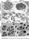
The PLCγ site of TrkB is involved in the maturation of nerve fibers. (A-D) Cross sections of P100 nerves to the posterior cristae of TrkbPLC/PLC and aged matched control mice are shown. (E-F) TEM images from P100 nerves to the posterior cristae; note that TrkbPLC/PLC mice show similar number of nerve fibers compared to the control animals, but nerve fibers tend to be much smaller in diameter with less myelin. Left brace "{" indicates the thickness of myelin in E-F panels and shows that reduction varies between fibers of comparable diameter of the neuronal process. Scale bars, 100 μm in A-D, 1 μm in E, F.
Figure 5
Figure 5
Afferent fibers are disorientated inside the vestibular sensory epithelia in absence of the TrkB/PLCγ docking site. (A-D) Shown are afferent fibers to the utricle labelled from the cerebellum; (A, C), P0 TrkbWT/WT control, and (B, D) P0 TrkbPLC/PLC point mutants. Note the highly focused projection in the control mice that already displays partial calyx formation (outlined by circles, A, C) with no fibers extending along the epithelial perimeters (dotted outline). In contrast, TrkbPLC/PLC mice show fibers extending for long distances along the epithelial perimeters (dotted outline) as well as within the sensory epithelium (arrows in panels B, D). There is only an occasional indication of partial calyx formation in TrkbPLC/PLC mutants at this stage (B, white circle). Scale bars, 100 μm in (A, B) and 10 μm in (C, D).
Figure 6
Figure 6
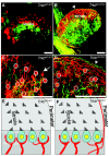
Abnormal calyx formation and hair cell innervations in absence of a functional TrkB/PLCγ site. (A-F) P8 utricular afferent projections labelled with lipophilic dye injected either into the cerebellum (red) or the brainstem (green) were compared. Note that the overall sorting of afferent fibers within the utricle is comparable between the TrkbPLC/PLC mutant mice and controls. However, many more fibers overlap in the TrkbPLC/PLC mutants compared to control (yellow in A, B). (C) Closer examination shows numerous hair cells surrounded by partial or complete calyces (indicated by dashed white circles in the striola region) in TrkbWT/WT controls, white circles indicate those near the edge of the striola region that are found only in TrkbWT/WT control mice. White arrow points to a peripheral calyx with nerve fiber coming off (so called mixed calyx/bouton fiber in the non-striola region). (D) Note absence of calyces outside the striola region in TrkbPLC/PLC mutants, and only occasional calyces inside the striola region of the TrkbPLC/PLC mutant mice (indicated by dashed white circles). Moreover, the long distances fibers are running inside the sensory epithelium as well as along the perimeter in the TrkbPLC/PLC mutant mice (arrowhead) (B, D), whereas much shorter and branched trajectories are typical in control mice (C). (E-F) Schematic drawings showing hair cell innervation within the striola region of the utricular sensory epithelium (E-F); the striola region is characterized by polarity reversal of hair cells and the presence of calyces around type 1 hair cells. Note that calyces in TrkbPLC/PLC mutants tend to be smaller, less frequent in the striola region and incomplete or absent near the perimeter (F); whereas TrkbWT/WT controls form calyces both inside and outside of the striola region (E). Moreover, unique to TrkbPLC/PLC mutants are afferent fibers that run along the perimeter of the sensory epithelia for long distance. Scale bars, 100 μm.
Figure 7
Figure 7
Histological evidence for the inability of calyx formation in TrkbPLC/PLC point mutants. (A) Adapted scheme showing major differences between type I and type II hair cells. (B-C) Light microscopy pictures showing the appearance of utricular sensory epithelia at postnatal day 100 with large calyceal spaces around type I hair cells in the TrkbWT/WT control (asterisks) (B), compared to little calyx formation in the TrkbPLC/PLC mutant mice (asterik) (C); arrows in both panel B and C indicate the stereocilia suggesting that hair cells are normally developed. Braces "{" indicate the supporting cells (SP), or the hair cells (HC). (D-E) Electron microscopy (EM) micrographs showing a typical type I hair cell in the utricle of TrkbPLC/PLC mutant mice mostly without a fully formed calyx in vivo. However, in the striola region there is an occasional type I hair cell with a fully formed calyx (E) in the utricle of TrkbPLC/PLC mutant mice. (F) A canal cristae type I hair cell of the TrkbPLC/PLC mutants innervated by several afferent only forming partial calices and efferent boutons (H). The existence of two types of hair cells is verified by the examination of hair cell apices, which show the presence of two different sizes of stereocilia and mitochondria but no calyx (G) as previously described [19,47]. Scale bars, 100 μm (panels B-C), 10 μm (panels D-F), (G-H are twice the magnification of F).
Figure 8
Figure 8
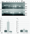
TRPC3 is upregulated in TrkbPLC/PLC mutants. (A-B) Trpc isoform expression analysis in mouse adult vestibular tissue. Expression analysis of Trpc1-7 isoforms was performed by RT-PCR using as control total RNA preparations from a pool (A) consisting of E15 brain and retina and dissected vestibular ganglion tissue (B). All PCR products were verified by direct sequence analyses. The negative control was a PCR reaction with the total RNA template without the addition of reverse transcriptase. A 1 kb ladder was used as a standard size marker (STD) and the corresponding kb lengths were indicated. (C) RT-PCR for Trpc3 showed increased expression of this channel in the vestibular organ of TrkbPLC/PLC mutants compared to both controls (wt and TrkbWT/WT, p = 0.02). No difference was detected between the two controls (p = 0.5). (D) Neuron specific enolase (NSE) was used as an internal control specific to neurons. No significant difference was observed between the TrkbPLC/PLC mutants and the two controls.
Figure 9
Figure 9
Calyx formation does not regulate expression patterns of KCNQ4. (A-D) Show the expression analysis of KCNQ4 in control (TrkbWT/WT) and TrkbPLC/PLC mutant mice at two different stages, P0 and P24. Upregulation of KCNQ4 protein shows only minor quantitative differences between control and TrkbPLC/PLC mutant. Scale bars, 100 μm.

Similar articles

Cited by

References

    1. Lysakowski AL, Goldberg JM. In: The Anatomy and Physiology of the Central and Peripheral Vestibular System. Highstein S, Popper AN, Fay RR, editor. New York: Springer; 2003. Morphophysiology of the vestibular periphery; pp. 57–152.
    1. Pirvola U, Ylikoski J, Palgi J, Lehtonen E, Arumae U, Saarma M. Brain-derived neurotrophic factor and neurotrophin 3 mRNAs in the peripheral target fields of developing inner ear ganglia. Proc Natl Acad Sci USA. 1992;89(20):9915–9919. doi: 10.1073/pnas.89.20.9915. - DOI - PMC - PubMed
    1. Spoendlin H. In: In Physiology of the ear. Jahn A, Santos-Sachi J, editor. New York: Raven Press; 1988. Neural anatomy of the inner ear; pp. 201–219.
    1. Fritzsch B, Tessarollo L, Coppola V, Reichardt LF. Neurotrophins in the ear: their roles in sensory neuron survival and fiber guidance. Prog Brain Res. 2004;146:265–278. full_text. - PubMed
    1. Minichiello L, Calella AM, Medina DL, Bonhoeffer T, Klein R, Korte M. Mechanism of TrkB-mediated hippocampal long-term potentiation. Neuron. 2002;36(1):121–137. doi: 10.1016/S0896-6273(02)00942-X. - DOI - PubMed

Publication types

MeSH terms

LinkOut - more resources