

Disrupted postnatal lung development in heme oxygenase-1 deficient mice
- PMID: 20932343
- PMCID: PMC2964616
- DOI: 10.1186/1465-9921-11-142
Disrupted postnatal lung development in heme oxygenase-1 deficient mice
Abstract
Background: Heme oxygenase (HO) degrades cellular heme to carbon monoxide, iron and biliverdin. The HO-1 isoform is both inducible and cyto-protective during oxidative stress, inflammation and lung injury. However, little is known about its precise role and function in lung development. We hypothesized that HO-1 is required for mouse postnatal lung alveolar development and that vascular expression of HO-1 is essential and protective during postnatal alveolar development.
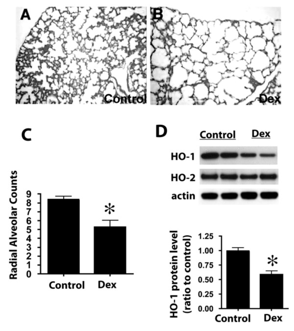
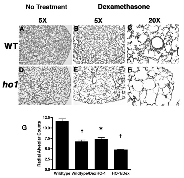
Methods: Neonatal lung development in wildtype and HO-1 mutant mice was evaluated by histological and molecular methods. Furthermore, these newborn mice were treated with postnatal dexamethasone (Dex) till postnatal 14 days, and evaluated for lung development.
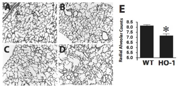
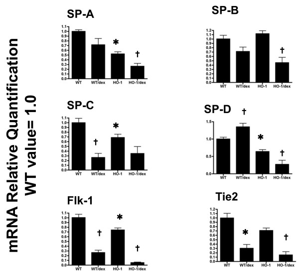
Results: Compared to wildtype littermates, HO-1 mutant mice exhibited disrupted lung alveolar structure including simplification, disorganization and reduced secondary crest formation. These defects in alveolar development were more pronounced when these mice were challenged with Dex treatment. Expression levels of both vascular endothelial and alveolar epithelial markers were also further decreased in HO-1 mutants after Dex treatment.
Conclusions: These experiments demonstrate that HO-1 is required in normal lung development and that HO-1 disruption and dexamethasone exposure are additive in the disruption of postnatal lung growth. We speculate that HO-1 is involved in postnatal lung development through modulation of pulmonary vascular development.
Figures
Similar articles
-
Vasculoprotective effects of heme oxygenase-1 in a murine model of hyperoxia-induced bronchopulmonary dysplasia.Am J Physiol Lung Cell Mol Physiol. 2012 Apr 15;302(8):L775-84. doi: 10.1152/ajplung.00196.2011. Epub 2012 Jan 27. Am J Physiol Lung Cell Mol Physiol. 2012. PMID: 22287607 Free PMC article.
-
Heme oxygenase-1 regulates postnatal lung repair after hyperoxia: role of β-catenin/hnRNPK signaling.Redox Biol. 2013 Feb 8;1(1):234-43. doi: 10.1016/j.redox.2013.01.013. eCollection 2013. Redox Biol. 2013. PMID: 24024157 Free PMC article.
-
MiR-196a regulates heme oxygenase-1 by silencing Bach1 in the neonatal mouse lung.Am J Physiol Lung Cell Mol Physiol. 2016 Aug 1;311(2):L400-11. doi: 10.1152/ajplung.00428.2015. Epub 2016 Jun 24. Am J Physiol Lung Cell Mol Physiol. 2016. PMID: 27343195 Free PMC article.
-
Heme oxygenase-1 and the vascular bed: from molecular mechanisms to therapeutic opportunities.Antioxid Redox Signal. 2008 Oct;10(10):1767-812. doi: 10.1089/ars.2008.2043. Antioxid Redox Signal. 2008. PMID: 18576916 Review.
-
The role of heme oxygenase-1 in pulmonary disease.Am J Respir Cell Mol Biol. 2007 Feb;36(2):158-65. doi: 10.1165/rcmb.2006-0331TR. Epub 2006 Sep 15. Am J Respir Cell Mol Biol. 2007. PMID: 16980551 Free PMC article. Review.
Cited by
-
Altered Expression of Heme Oxygenase 2 in Heme Oxygenase 1-deficient Mouse Embryos.J Histochem Cytochem. 2023 Aug;71(8):431-450. doi: 10.1369/00221554231189310. Epub 2023 Jul 22. J Histochem Cytochem. 2023. PMID: 37480265 Free PMC article.
-
Plasticity of button-like junctions in the endothelium of airway lymphatics in development and inflammation.Am J Pathol. 2012 Jun;180(6):2561-75. doi: 10.1016/j.ajpath.2012.02.019. Epub 2012 Apr 23. Am J Pathol. 2012. PMID: 22538088 Free PMC article.
-
Two Faces of Heme Catabolic Pathway in Newborns: A Potential Role of Bilirubin and Carbon Monoxide in Neonatal Inflammatory Diseases.Oxid Med Cell Longev. 2020 Aug 18;2020:7140496. doi: 10.1155/2020/7140496. eCollection 2020. Oxid Med Cell Longev. 2020. PMID: 32908636 Free PMC article. Review.
-
The Nuclear Translocation of Heme Oxygenase-1 in Human Diseases.Front Cell Dev Biol. 2022 Jun 29;10:890186. doi: 10.3389/fcell.2022.890186. eCollection 2022. Front Cell Dev Biol. 2022. PMID: 35846361 Free PMC article. Review.
-
Induction of heme oxygenase-1 by hemin protects lung against orthotopic autologous liver transplantation-induced acute lung injury in rats.J Transl Med. 2016 Feb 2;14:35. doi: 10.1186/s12967-016-0793-0. J Transl Med. 2016. PMID: 26838179 Free PMC article.
References
-
- Warner BB, Stuart LA, Papes RA, Wispe JR. Functional and pathological effects of prolonged hyperoxia in neonatal mice. Am J Physiol. 1998;275(1 Pt 1):L110–117. - PubMed
-
- Wagenaar GT, ter Horst SA, van Gastelen MA, Leijser LM, Mauad T, van der Velden PA, de Heer E, Hiemstra PS, Poorthuis BJ, Walther FJ. Gene expression profile and histopathology of experimental bronchopulmonary dysplasia induced by prolonged oxidative stress. Free Radic Biol Med. 2004;36(6):782–801. doi: 10.1016/j.freeradbiomed.2003.12.007. - DOI - PubMed
-
- Massaro D, Massaro GD. Dexamethasone accelerates postnatal alveolar wall thinning and alters wall composition. Am J Physiol. 1986;251(2 Pt 2):R218–224. - PubMed
Publication types
MeSH terms
Substances
Grants and funding
LinkOut - more resources
Full Text Sources
