

Specific antibody responses against membrane proteins of erythrocytes infected by Plasmodium falciparum of individuals briefly exposed to malaria
- PMID: 20932351
- PMCID: PMC2959075
- DOI: 10.1186/1475-2875-9-276
Specific antibody responses against membrane proteins of erythrocytes infected by Plasmodium falciparum of individuals briefly exposed to malaria
Abstract
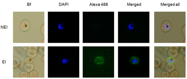
Background: Plasmodium falciparum infections could lead to severe malaria, principally in non-immune individuals as children and travellers from countries exempted of malaria. Severe malaria is often associated with the sequestration of P. falciparum-infected erythrocytes in deep micro-vascular beds via interactions between host endothelial receptors and parasite ligands expressed on the surface of the infected erythrocyte. Although, serological responses from individuals living in endemic areas against proteins expressed at surface of the infected erythrocyte have been largely studied, seldom data are available about the specific targets of antibody response from travellers.
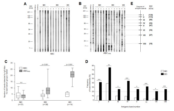
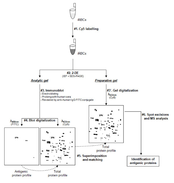
Methods: In order to characterize antigens recognized by traveller sera, a comparison of IgG immune response against membrane protein extracts from uninfected and P. falciparum-infected red blood cells (iRBC), using immunoblots, was performed between non exposed individuals (n = 31) and briefly exposed individuals (BEI) (n = 38) to malaria transmission.
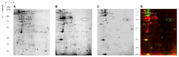
Results: Immune profile analysis indicated that eight protein bands from iRBC were significantly detected more frequently in the BEI group. Some of these antigenic proteins were identified by an original immuno-proteomic approach.
Conclusion: Collectively, these data may be useful to characterize the singular serological immune response against a primary malaria infection in individuals briefly exposed to transmission.
Figures
Similar articles
-
Antibodies from malaria-exposed pregnant women recognize trypsin resistant epitopes on the surface of Plasmodium falciparum-infected erythrocytes selected for adhesion to chondroitin sulphate A.Malar J. 2004 Sep 6;3:31. doi: 10.1186/1475-2875-3-31. Malar J. 2004. PMID: 15350207 Free PMC article.
-
Relationship of binding of immunoglobulin G to Plasmodium falciparum-infected erythrocytes with parasite endemicity and antibody responses to conserved antigen in immune individuals.Clin Diagn Lab Immunol. 2004 Jan;11(1):6-11. doi: 10.1128/cdli.11.1.6-11.2004. Clin Diagn Lab Immunol. 2004. PMID: 14715538 Free PMC article.
-
Comparative analysis of the profiles of IgG subclass-specific responses to Plasmodium falciparum apical membrane antigen-1 and merozoite surface protein-1 in naturally exposed individuals living in malaria hypoendemic settings, Iran.Malar J. 2015 Feb 5;14:58. doi: 10.1186/s12936-015-0547-0. Malar J. 2015. PMID: 25652589 Free PMC article.
-
PfEMP1 - A Parasite Protein Family of Key Importance in Plasmodium falciparum Malaria Immunity and Pathogenesis.Adv Parasitol. 2015 Apr;88:51-84. doi: 10.1016/bs.apar.2015.02.004. Epub 2015 Mar 23. Adv Parasitol. 2015. PMID: 25911365 Review.
-
VAR2CSA-Mediated Host Defense Evasion of Plasmodium falciparum Infected Erythrocytes in Placental Malaria.Front Immunol. 2021 Feb 9;11:624126. doi: 10.3389/fimmu.2020.624126. eCollection 2020. Front Immunol. 2021. PMID: 33633743 Free PMC article. Review.
Cited by
-
An Optimized Fluorescence-Based Bidimensional Immunoproteomic Approach for Accurate Screening of Autoantibodies.PLoS One. 2015 Jul 1;10(7):e0132142. doi: 10.1371/journal.pone.0132142. eCollection 2015. PLoS One. 2015. PMID: 26132557 Free PMC article.
-
Altered protein networks and cellular pathways in severe west nile disease in mice.PLoS One. 2013 Jul 10;8(7):e68318. doi: 10.1371/journal.pone.0068318. Print 2013. PLoS One. 2013. PMID: 23874584 Free PMC article.
-
Naturally Acquired Antibodies against Plasmodium falciparum: Friend or Foe?Pathogens. 2021 Jul 2;10(7):832. doi: 10.3390/pathogens10070832. Pathogens. 2021. PMID: 34357982 Free PMC article. Review.
-
The Identification of Native Epitopes Eliciting a Protective High-Affinity Immunoglobulin Subclass Response to Blood Stages of Plasmodium falciparum: Protocol for Observational Studies.JMIR Res Protoc. 2020 Jul 17;9(7):e15690. doi: 10.2196/15690. JMIR Res Protoc. 2020. PMID: 32706743 Free PMC article.
-
Erythrocyte membrane proteins involved in the immune response to Plasmodium falciparum and Plasmodium vivax infection.Parasitol Res. 2021 May;120(5):1789-1797. doi: 10.1007/s00436-021-07135-6. Epub 2021 Apr 2. Parasitol Res. 2021. PMID: 33797613
References
Publication types
MeSH terms
Substances
LinkOut - more resources
Full Text Sources
Other Literature Sources
