Review
. 2011 Jun 15;69(12):e55-68.
doi: 10.1016/j.biopsych.2010.07.024. Epub 2010 Oct 8.

From reactive to proactive and selective control: developing a richer model for stopping inappropriate responses

Affiliations
Review

From reactive to proactive and selective control: developing a richer model for stopping inappropriate responses

Adam R Aron. Biol Psychiatry. .

Abstract

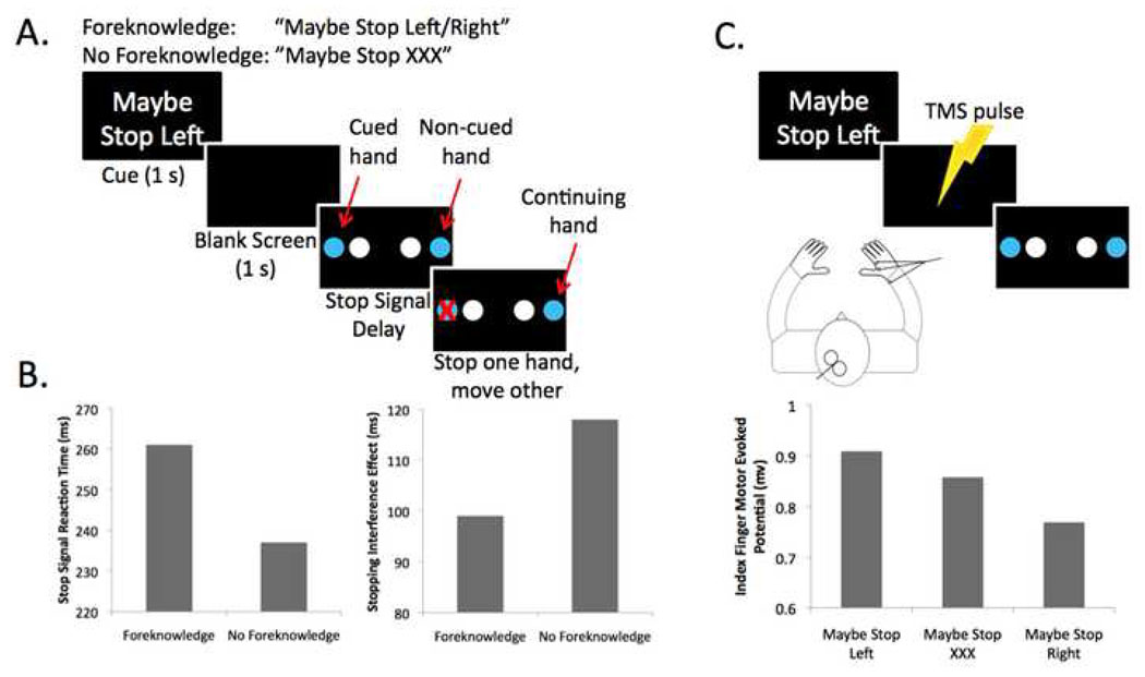
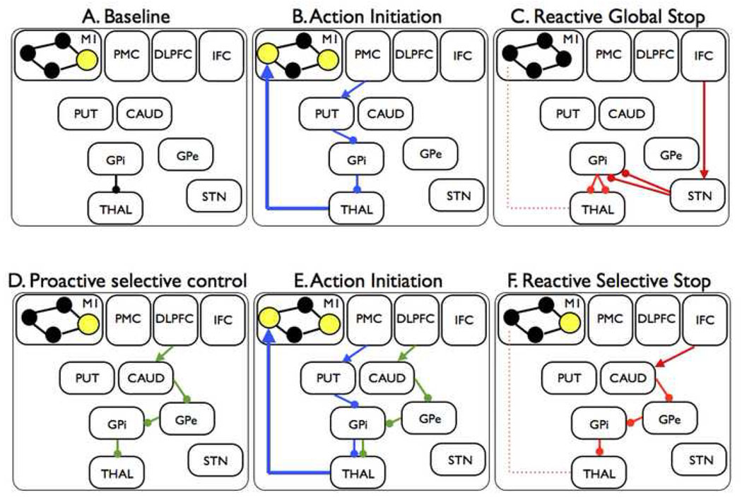
A better understanding of the neural systems underlying impulse control is important for psychiatry. Although most impulses are motivational or emotional rather than motoric per se, it is research into the neural architecture of motor response control that has made the greatest strides. This article reviews recent developments in the cognitive neuroscience of stopping responses. Most research of this kind has focused on reactive control-that is, how subjects stop a response outright when instructed by a signal. It is argued that reactive paradigms are limited as models of control relevant to psychiatry. Instead, a set of paradigms is advocated that begins to model proactive inhibitory control-that is, how a subject prepares to stop an upcoming response tendency. Proactive inhibitory control is generated according to the goals of the subject rather than by an external signal, and it can be selectively targeted at a particular response tendency. This may have wider validity than reactive control as an experimental model for stopping inappropriate responses.


Conflict of interest statement

Financial Disclosure

The author reports no biomedical financial interests or potential conflicts of interest.

Figures

Figure 1
Figure 1
Three behavioral paradigms for measuring stopping. A. The stop signal test. A ready signal (small box) is presented followed by a Go signal (left-pointing arrow). The subject initiates and executes a left button press (Go trial). The next trial begins in the same way, however a stop signal (auditory) is presented at a delay (e.g. 200 ms) after the Go signal. The subject stops the response (stop trial). B. The Go/NoGo test. Typically this is done with a stream of letters. The subject responds to all except the letter 'X'. C. The antisaccade test. The central eye fixation signal is a colored box. Here, green means 'make a saccade' in the direction of the upcoming target, while blue means 'make a saccade in the opposite direction to the upcoming target'. On the antisaccade trial shown above, the target triggers an automatic saccade to the left, but the subject over-rides this to move her eyes to the right.
Figure 2
Figure 2
Functional MRI studies of stop signal and related paradigms. The ‘stopping network’ in the cortex is activated by different control tasks and is predominantly right-lateralized. It includes the presupplementary motor area (preSMA) and the right inferior frontal cortex (rIFC). Right IFC activity is broadly distributed and may reflect an inferior frontal junction (IFJ) component, a more ventral posterior inferior frontal (pIFG) (putatively implementing inhibitory control) and an insula region of unknown function. Maps of the activation during performance of go/no-go, stop-signal and antisaccade tasks were revealed by contrasting no-go vs. frequent-go, stop vs. go, and antisaccade vs. baseline-saccade trials, respectively, see (16) for further details. Copyright permission requested.
Figure 3
Figure 3
The brain network for reactive stopping. A. Regions that are critical for stopping in the standard stop signal paradigm. Two regions within the inferior frontal cortex (IFC) are the inferior frontal junction (IFJ) and the posterior inferior frontal gyrus (pIFG). The presupplementary motor area (preSMA) is in the medial surface. B. White-matter tractography using diffusion tensor imaging reveals a three-way network in the right hemisphere between nodes that are critical for stopping action (31). Copyright permission requested.
Figure 4
Figure 4
Neurophysiological recording from the striatum shows the signature of proactive inhibitory control. A. Monkeys were studied with a symmetrically reinforced delayed Go/NoGo task (88). The trial began with a lever press. A Go or NoGo cue was then presented. After an interval of 2.5 to 3.5 secs, a trigger stimulus was presented, requiring the animal to either release the lever or not release it to get reward. B. Some neurons in the striatum showed a pattern of sustained activity between the NoGo instrution and the trigger stimulus (perhaps involved in preparing to inhibit movement) C. Other neurons showed sustained activity between the trigger stimulus and the reward (perhaps implementing the movement inhibition itself). Copyright permission requested.
Figure 5
Figure 5
Behavioral and TMS studies of selective stopping. A. The selective stopping task has stopping goal and no stopping goal conditions. Each trial begins with a cue providing a stopping goal (foreknowledge) or none (no foreknowledge). This was followed by a blank screen. A Go stimulus was then presented, requiring the subject to initiate a bimanual response with index fingers or middle fingers of each hand. On a minority of trials, a visual stop signal (red ‘X’) occurred and the subject tried to stop the indicated hand, while continuing with the other hand. B. When a stopping goal is provided (foreknowledge), the speed of stopping, SSRT, is longer, while the degree of slowing of the continuing hand is less (79). Taken together this pattern of data suggests that stopping without a stopping goal (or with less information about what to stop) may recruit a mechanism with more global effects, but which is also quicker. C. For the same paradigm, TMS was delivered to left M1 with electromyography recorded from the right hand. The level of corticomotor excitability was significantly less when subjects anticipated they would have to stop the right hand – consistent with the possibility of proactive selective inhibitory control (126).
Figure 6
Figure 6
Hypothetical fronto-basal-ganglia circuits for global and selective stopping. A. When the subject's hand is at rest, the GPi is tonically inhibiting thalamocortical output to hand representations so that these are only weakly active (small filled circles). By contrast, one M1 representation, for example for the speech system, is strongly active (large yellow-filled circle). B. The subject initiates a hand movement using the direct pathway. The PMC activates the putamen, the putamen inhibits the GPi, this removes inhibition from the thalamus and increases drive to the hand area of M1. C. The IFC sends input to the STN via the hyperdirect pathway. The STN has a broad effect on GPi, leading to global suppression of thalamocortical programs, including hand and speech systems. D. Proactive selective control may be set up via the indirect pathway. The DLPFC activates a specific channel of the caudate, the caudate inhibits a specific channel of the GPe, the GPe inhibits a specific channel of the GPi (directly or via the STN) and inhibition of a particular thalamocortical channel is prepared (but perhaps not triggered until stopping is needed). E. Action initiation occurs as for B. above, except it occurs with the proactive selective control system activated. This could lead to slower response emission. F. The indirect pathway may be triggered by the IFC when a stop signal occurs. This leads to suppression of one, but not all, representations in M1. M1 = primary motor cortex; PMC = premotor cortex; DLPFC = dorsolateral prefrontal cortex; IFC = inferior frontal cortex; PUT = putamen; CAUD = caudate; GPi = globus pallidus pars interna; GPe = globus pallidus pars externa; STN = subthalamic nucleus; THAL = thalamus.

References

    1. Gottesman II, Gould TD. The endophenotype concept in psychiatry: etymology and strategic intentions. Am J Psychiatry. 2003;160:636–645. - PubMed
    1. Goldin PR, McRae K, Ramel W, Gross JJ. The neural bases of emotion regulation: reappraisal and suppression of negative emotion. Biol Psychiatry. 2008;63:577–586. - PMC - PubMed
    1. Ochsner KN, Gross JJ. The cognitive control of emotion. Trends Cogn Sci. 2005;9:242–249. - PubMed
    1. Banich MT, Mackiewicz KL, Depue BE, Whitmer AJ, Miller GA, Heller W. Cognitive control mechanisms, emotion and memory: a neural perspective with implications for psychopathology. Neurosci Biobehav Rev. 2009;33:613–630. - PMC - PubMed
    1. Brody AL, Mandelkern MA, Olmstead RE, Jou J, Tiongson E, Allen V, et al. Neural substrates of resisting craving during cigarette cue exposure. Biol Psychiatry. 2007;62:642–651. - PMC - PubMed

Publication types