Review
. 2011 Jan;32(1):70-83.
doi: 10.1016/j.yfrne.2010.09.001. Epub 2010 Oct 25.

New aspects of melanocortin signaling: a role for PRCP in α-MSH degradation

Affiliations
Review

New aspects of melanocortin signaling: a role for PRCP in α-MSH degradation

Sabrina Diano. Front Neuroendocrinol. 2011 Jan.

Abstract

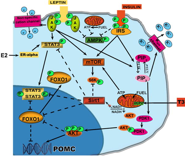
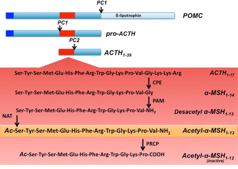
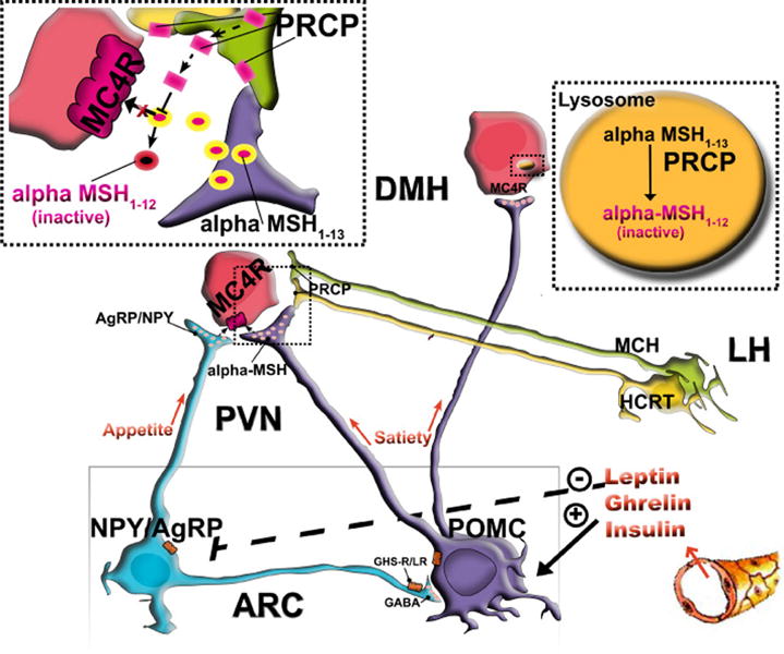
The role of the central melanocortin system in the regulation of energy metabolism has received much attention during the past decade since gene mutations of key components in melanocortin signaling cause monogenic forms of obesity in animals and humans. In the arcuate nucleus of the hypothalamus the prohormone proopiomelanocortin (POMC) is posttranslationally cleaved to produce α-melanocyte stimulating hormone (α-MSH), a peptide with anorexigenic effects upon activation of the melanocortin receptors (MCRs). α-MSH undergoes extensive post-translational processing and its in vivo activity is short lived due to rapid degradation. The enzymatic process that controls α-MSH inactivation is incompletely understood. Recent evidence suggests that prolyl carboxypeptidase (PRCP) is an enzyme responsible for α-MSH degradation. As for many key melanocortin peptides, gene mutation of PRCP causes a change in the metabolic phenotype of rodents. This review summarizes the current knowledge on the melanocortin system with particular focus on PRCP, a newly discovered component of the melanocortin system.


Figures

Figure 1
Figure 1
Schematic illustration showing the metabolic pathways activated by hormones and fuel availability in POMC neurons of the arcuate nucleus of the hypothalamus. Activation of PI3K in POMC neurons by either leptin or insulin receptors induces activation of AKT which leads to the phosphorylation of FoxO1 and thus its exclusion from the nucleus allowing the transcription of POMC. Concomitantly, PI3K activation also induces the opening of fewer potassium ATP channels thus inducing the leptin-dependent firing of the POMC neurons and leptin-dependent POMC transcription via Stat3. Stat3 has been also reported to be a target of estrogen signaling. However, the effect of estrogen on Stat3 is JAK independent. In satiety conditions when POMC neurons are activated, Sirt1 and NAD+ levels are decreased and Sirt1 activation of FoxO1 is also inhibited. Finally, the decrease in the ratio AMP/ATP will decrease AMPK while increase mTOR activity.
Figure 2
Figure 2
Schematic illustration of hypothalamic POMC processing. Prohormone convertase 1 (PC1) cleaves the 32 kDa preprotein into proACTH and β-lipotrophin . ProACTH is then further converted by PC1 to form ACTH1–39. Prohormone convertase 2 (PC2) will then cleave ACTH1–39, to form ACTH1–17. ACTH1–17 will be first processed by carboxypeptidase E (CPE) and then α-amidating mono-oxygenase (PAM) to form desacetyl α-MSH1–13. An n-actelyltransferase (NAT) will then convert desacetyl-α-MSH1–13 into the more active form acetyl-α-MSH1–13. Finally acetyl- α-MSH1–13 will be degraded by prolyl carboxypeptidase (PRCP) into the inactive product acetyl- α-MSH1–12.
Figure 3
Figure 3
PRCP is in an anatomical position to determine the efficacy of released α-MSH, thus controlling the output of the melanocortin system. PRCP is mainly expressed in the lateral hypothalamus (LH) in hypocretin/orexin (Hcrt) and melanin-concentrating hormone (MCH) neurons and in the hypothalamic dorsomedial nucleus (DMH) where MC4R-expressing neurons are located. The Hcrt and MCH neurons project to various areas of the hypothalamus, such as the paraventricular nucleus, where α-MSH terminals strongly innervate MC4R-expressing neurons. When released from the Hcrt and/or MCH terminals, PRCP will degrade α-MSH extracellularly, thus increasing the antagonistic effect of AgRP. In the DMH, however, PRCP degrades α-MSH intracellularly, thereby enhancing the overall orexigenic tone of the system.

References

    1. Ainscow EK, et al. Dynamic imaging of free cytosolic ATP concentration during fuel sensing by rat hypothalamic neurones: evidence for ATP-independent control of ATP-sensitive K(+) channels. J Physiol. 2002;544:429–45. - PMC - PubMed
    1. Ainslie DA, Morris MJ, Wittert G, Turnbull H, Proietto J, Thorburn AW. Estrogen deficiency causes central leptin insensitivity and increased hypothalamic neuropeptide Y. Int J Obes Relat Metab Disord. 2001;25:1680–8. - PubMed
    1. Andrews ZB, Liu ZW, Walllingford N, Erion DM, Borok E, Friedman JM, Tschöp MH, Shanabrough M, Cline G, Shulman GI, Coppola A, Gao XB, Horvath TL, Diano S. Uncoupling protein-2 mediates ghrelin’s action on NPY/AgRP neurons. Nature. 2008;454:846–51. - PMC - PubMed
    1. Bady I, Marty N, Dallaporta M, Emery M, Gyger J, Tarussio D, Foretz M, Thorens B. Evidence from glut2-null mice that glucose is a critical physiological regulator of feeding. Diabetes. 2006;55:988–95. - PubMed
    1. Bagnasco M, Dube MG, Kalra PS, Kalra SP. Evidence for the existence of distinct central appetite, energy expenditure, and ghrelin stimulation pathways as revealed by hypothalamic site-specific leptin gene therapy. Endocrinology. 2002;143:4409–4421. - PubMed

Publication types

MeSH terms