Review
. 2011 Oct;1812(10):1281-90.
doi: 10.1016/j.bbadis.2010.09.016. Epub 2010 Oct 12.

Calcium-mediated mechanisms of cystic expansion

Affiliations
Review

Calcium-mediated mechanisms of cystic expansion

Shakila Abdul-Majeed et al. Biochim Biophys Acta. 2011 Oct.

Abstract

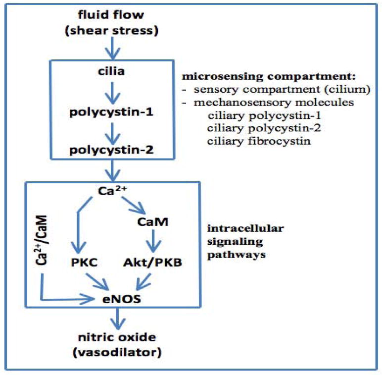
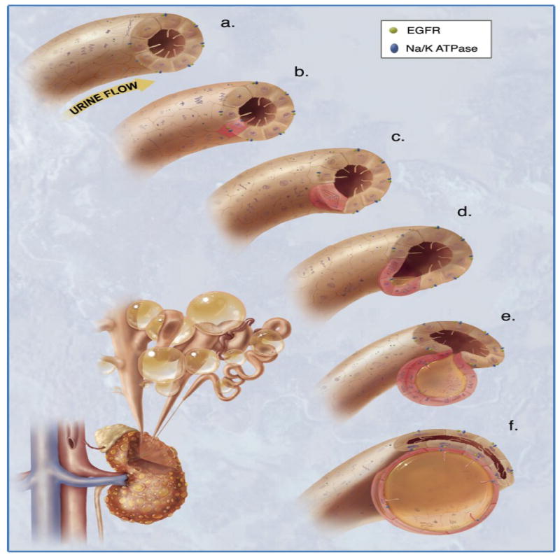
In this review, we will discuss several well-accepted signaling pathways toward calcium-mediated mechanisms of cystic expansion. The second messenger calcium ion has contributed to a vast diversity of signal transduction pathways. We will dissect calcium signaling as a possible mechanism that contributes to renal cyst formation. Because cytosolic calcium also regulates an array of signaling pathways, we will first discuss cilia-induced calcium fluxes, followed by Wnt signaling that has attributed to much-discussed planar cell polarity. We will then look at the relationship between cytosolic calcium and cAMP as one of the most important aspects of cyst progression. The signaling of cAMP on MAPK and mTOR will also be discussed. We infer that while cilia-induced calcium fluxes may be the initial signaling messenger for various cellular pathways, no single signaling mediator or pathway is implicated exclusively in the progression of the cystic expansion. This article is part of a Special Issue entitled: Polycystic Kidney Disease.


Figures

Figure 1
Figure 1. Cysto-protein complex
Polycystin-1 and polycystin-2 form a complex with each other at their COOH termini. Functioning as an adaptor protein, Kif bridges the interaction of between fibrocystin and polycystin complex. Figure is reproduced from Kolb, et al with permission [39].
Figure 2
Figure 2. Primary cilia in signal transduction system
Cilia sense fluid-shear stress on the apical membrane of the cells. Fluid flow that produces enough drag-force on the top of the cells will bend sensory cilia. Bending of cilia will activate the cysto-proteins, resulting in extracellular calcium influx. Figure is reproduced from AbouAlaiwi, et al with permission [38].
Figure 3
Figure 3. Mechanosensory cilia, cytosolic calcium and nitric oxide production
Bending or activation of the cilia involves mechanosensory polycystin-1 and polycystin-2 complex, which results in biochemical synthesis of NO. The signaling pathway requires extracellular calcium influx (Ca2+), followed by activation of various calcium-dependent proteins including calmodulin (CaM) and protein kinase C (PKC). Together with PKB, CaM and PKC are important downstream molecular components to activate endothelial nitric oxide synthase (eNOS).
Figure 4
Figure 4. Mechanosensory cilia, cytosolic calcium and Wnt signaling
Bending or activation of the cilia results in maintaining cytosolic calcium (Ca2+), followed by activation of inversin. Calcium and inversin function as the molecular switches for Wnt signaling pathways. The Wnt canonical pathway involves β-catenin. Both β-catenin and E-cadherin regulate cell differentiation and proliferation. The Wnt non-canonical pathway involves small GTPase rho and JNK, both of which play an important role in planar cell polarity.
Figure 5
Figure 5. Planar cell polarity and cystic kidney disease
The illustration depicts the mechanism of mitotic spindle formation as an index of planar cell polarity in cyst expansion. a. Each cilium senses changes in urine flow. This message provides critical calcium signals to the cell. b. Abnormalities in any of the cysto-proteins results in abnormal ciliary function. c. The functional abnormality in ciliary sensing results in disorientation of the mitotic spindle, and hence the cell will lose planar cell polarity. d. Direction of cell division becomes randomized. This will result in increasing tubular diameter, rather than tubular elongation. e. Budding of a cyst from the renal tubule occurs. The abnormal localizations of epidermal growth factor receptor (EGFR) and Na+/K+ ATPase pump are typical characteristics of polycystic kidneys. f. The cyst is eventually enlarged and expanded further. Multiple cysts within a kidney are illustrated on the bottom left corner. Figure is reproduced from Kolb, et al with permission [39].
Figure 6
Figure 6. Cytosolic calcium and cAMP signaling
Under normal conditions, the carboxy terminal tails of polycystin-1 and polycystin-2 interact, which enables calcium entry into the cell. This further stimulates the release of calcium from the calcium stores inside the cell. In the presence of high, steady state levels of calcium, calcium-stimulated phosphodiesterases are capable of degrading cAMP into AMP, thereby controlling the cAMP levels in the cell. The low levels of cAMP are unable to stimulate MAPK pathway. In polycystic kidneys, however, the resulting abnormality in cysto-proteins causes a lower level of intracellular calcium. Hence, the activity of phosphodiesterases is not stimulated and cAMP level increases, leading to activation of B-Raf of MAPK pathway.
Figure 7
Figure 7. Signaling pathway of mTOR
In healthy kidney cells, the cysto-protein complex interacts with tuberin. Hamartin and tuberin form a complex and localize in the basal body. Tuberin is a GTPase activating protein (GAP) which controls the activity of Rheb. Tuberin inactivates Rheb, thereby inhibiting the mTOR pathway. In polycystic kidneys, however, tuberin is no longer protected in this complex and is phosphorylated by Akt, RSK (via ERK). As a result, tubulin is unable to form a heteradimer with hamartin. Hence, tuberin is no longer able inhibit Rheb, and the mTOR pathway is activated.

Similar articles

Cited by

References

    1. Polycystic kidney disease: the complete structure of the PKD1 gene and its protein. The International Polycystic Kidney Disease Consortium, Cell. 1995;81:289–298. - PubMed
    1. Mochizuki T, Wu G, Hayashi T, Xenophontos SL, Veldhuisen B, Saris JJ, Reynolds DM, Cai Y, Gabow PA, Pierides A, Kimberling WJ, Breuning MH, Deltas CC, Peters DJ, Somlo S. PKD2, a gene for polycystic kidney disease that encodes an integral membrane protein, Science. Vol. 272. New York, N.Y: 1996. pp. 1339–1342. - PubMed
    1. Ward CJ, Hogan MC, Rossetti S, Walker D, Sneddon T, Wang X, Kubly V, Cunningham JM, Bacallao R, Ishibashi M, Milliner DS, Torres VE, Harris PC. The gene mutated in autosomal recessive polycystic kidney disease encodes a large, receptor-like protein. Nature genetics. 2002;30:259–269. - PubMed
    1. Cai Y, Maeda Y, Cedzich A, Torres VE, Wu G, Hayashi T, Mochizuki T, Park JH, Witzgall R, Somlo S. Identification and characterization of polycystin-2, the PKD2 gene product. The Journal of biological chemistry. 1999;274:28557–28565. - PubMed
    1. Delmas P, Nomura H, Li X, Lakkis M, Luo Y, Segal Y, Fernandez-Fernandez JM, Harris P, Frischauf AM, Brown DA, Zhou J. Constitutive activation of G-proteins by polycystin-1 is antagonized by polycystin-2. The Journal of biological chemistry. 2002;277:11276–11283. - PubMed

Publication types

MeSH terms

LinkOut - more resources