

Reducing the gradient artefact in simultaneous EEG-fMRI by adjusting the subject's axial position
- PMID: 20932913
- PMCID: PMC3095086
- DOI: 10.1016/j.neuroimage.2010.09.079
Reducing the gradient artefact in simultaneous EEG-fMRI by adjusting the subject's axial position
Abstract
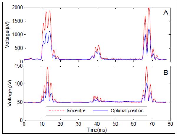
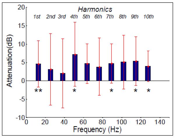
Large artefacts that compromise EEG data quality are generated when electroencephalography (EEG) and functional magnetic resonance imaging (fMRI) are carried out concurrently. The gradient artefact produced by the time-varying magnetic field gradients is the largest of these artefacts. Although average artefact correction (AAS) and related techniques can remove the majority of this artefact, the need to avoid amplifier saturation necessitates the use of a large dynamic range and strong low-pass filtering in EEG recording. Any intrinsic reduction in the gradient artefact amplitude would allow data with a higher bandwidth to be acquired without amplifier saturation, thus increasing the frequency range of neuronal activity that can be investigated using combined EEG-fMRI. Furthermore, gradient artefact correction methods assume a constant artefact morphology over time, so their performance is compromised by subject movement. Since the resulting, residual gradient artefacts can easily swamp signals from brain activity, any reduction in their amplitude would be highly advantageous for simultaneous EEG-fMRI studies. The aim of this work was to investigate whether adjustment of the subject's axial position in the MRI scanner can reduce the amplitude of the induced gradient artefact, before and after artefact correction using AAS. The variation in gradient artefact amplitude as a function of the subject's axial position was first investigated in six subjects by applying gradient pulses along the three Cartesian axes. The results of this study showed that a significant reduction in the gradient artefact magnitude can be achieved by shifting the subject axially by 4 cm towards the feet relative to the standard subject position (nasion at iso-centre). In a further study, the 4-cm shift was shown to produce a 40% reduction in the RMS amplitude (and a 31% reduction in the range) of the gradient artefact generated during the execution of a standard multi-slice, EPI sequence. By picking out signals occurring at harmonics of the slice acquisition frequency, it was also shown that the 4-cm shift led to a 36% reduction in the residual gradient artefact after AAS. Functional and anatomical MR data quality is not affected by the 4-cm shift, as the head remains in the homogeneous region of the static magnet field and gradients.
Copyright © 2010 Elsevier Inc. All rights reserved.
Figures
Similar articles
-
Reference layer artefact subtraction (RLAS): a novel method of minimizing EEG artefacts during simultaneous fMRI.Neuroimage. 2014 Jan 1;84:307-19. doi: 10.1016/j.neuroimage.2013.08.039. Epub 2013 Aug 28. Neuroimage. 2014. PMID: 23994127
-
Understanding gradient artefacts in simultaneous EEG/fMRI.Neuroimage. 2009 Jun;46(2):459-71. doi: 10.1016/j.neuroimage.2009.01.029. Neuroimage. 2009. PMID: 19385014
-
Exploring the origins of EEG motion artefacts during simultaneous fMRI acquisition: Implications for motion artefact correction.Neuroimage. 2018 Jun;173:188-198. doi: 10.1016/j.neuroimage.2018.02.034. Epub 2018 Feb 25. Neuroimage. 2018. PMID: 29486322 Free PMC article.
-
Novel artefact removal algorithms for co-registered EEG/fMRI based on selective averaging and subtraction.Neuroimage. 2013 Jan 1;64:407-15. doi: 10.1016/j.neuroimage.2012.09.022. Epub 2012 Sep 17. Neuroimage. 2013. PMID: 22995780
-
Towards motion insensitive EEG-fMRI: Correcting motion-induced voltages and gradient artefact instability in EEG using an fMRI prospective motion correction (PMC) system.Neuroimage. 2016 Sep;138:13-27. doi: 10.1016/j.neuroimage.2016.05.003. Epub 2016 May 6. Neuroimage. 2016. PMID: 27157789
Cited by
-
An open-access dataset of naturalistic viewing using simultaneous EEG-fMRI.Sci Data. 2023 Aug 23;10(1):554. doi: 10.1038/s41597-023-02458-8. Sci Data. 2023. PMID: 37612297 Free PMC article.
-
Gradient Artefact Correction and Evaluation of the EEG Recorded Simultaneously with fMRI Data Using Optimised Moving-Average.J Med Eng. 2016;2016:9614323. doi: 10.1155/2016/9614323. Epub 2016 Jun 28. J Med Eng. 2016. PMID: 27446943 Free PMC article.
-
Two spatiotemporally distinct value systems shape reward-based learning in the human brain.Nat Commun. 2015 Sep 8;6:8107. doi: 10.1038/ncomms9107. Nat Commun. 2015. PMID: 26348160 Free PMC article.
-
Neural correlates of evidence accumulation during value-based decisions revealed via simultaneous EEG-fMRI.Nat Commun. 2017 Jun 9;8:15808. doi: 10.1038/ncomms15808. Nat Commun. 2017. PMID: 28598432 Free PMC article.
-
Interhemispheric Connectivity Supports Load-Dependent Working Memory Maintenance for Complex Visual Stimuli.Brain Connect. 2022 Dec;12(10):892-904. doi: 10.1089/brain.2021.0171. Epub 2022 Jun 1. Brain Connect. 2022. PMID: 35473394 Free PMC article.
References
-
- Allen PJ, Josephs O, Turner R. A Method for removing Imaging Artifact from Continuous EEG Recorded during Functional MRI. Neuroimage. 2000;12(2):230–239. - PubMed
-
- Allen PJ, Poizzi G, Krakow K, Fish DR, Lemieux L. Identification of EEG Events in the MR Scanner: The Problem of Pulse Artifact and a Method for Its Subtraction. Neuroimage. 1998;8(3):229–239. - PubMed
-
- Anami K, Mori T, Tanaka F, Kawagoe Y, Okamoto J, Yarita M, Ohnishi T, Yumoto M, Matsuda H, Saitoh O. Stepping stone sampling for retrieving artifact-free electoencephalogram during functional magnetic resonance imaging. Neuroimage. 2003;19(2):281–295. - PubMed
-
- Benar CG, Schon D, Grimault S, Nazarian B, Burle B, Roth M, Badier J-M, Marquis P, Liegeois-Chauvel C, Anton J-L. Single-Trial Analysis of Oddball Event-Related Potentials in Simultaneous EEG-fMRI. Human Brain Mapping. 2007;28:602–613. - PubMed
Publication types
MeSH terms
Grants and funding
LinkOut - more resources
Full Text Sources
Medical
