Review
. 2011 Jan 15;505(2):131-43.
doi: 10.1016/j.abb.2010.09.028. Epub 2010 Oct 7.

Enzymes of the mevalonate pathway of isoprenoid biosynthesis

Affiliations
Review

Enzymes of the mevalonate pathway of isoprenoid biosynthesis

Henry M Miziorko. Arch Biochem Biophys. .

Abstract

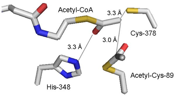
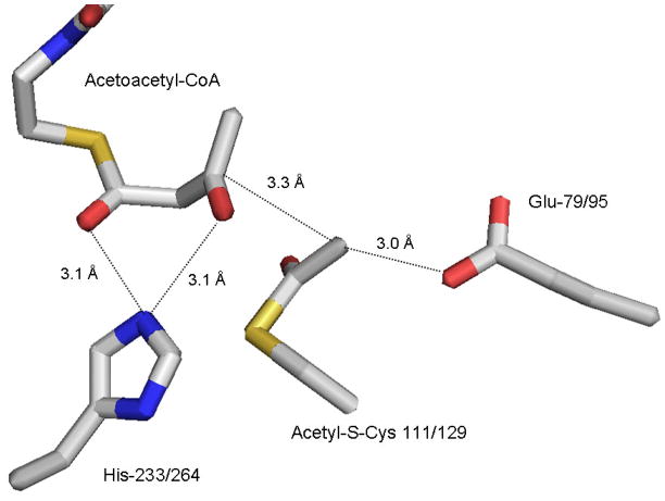
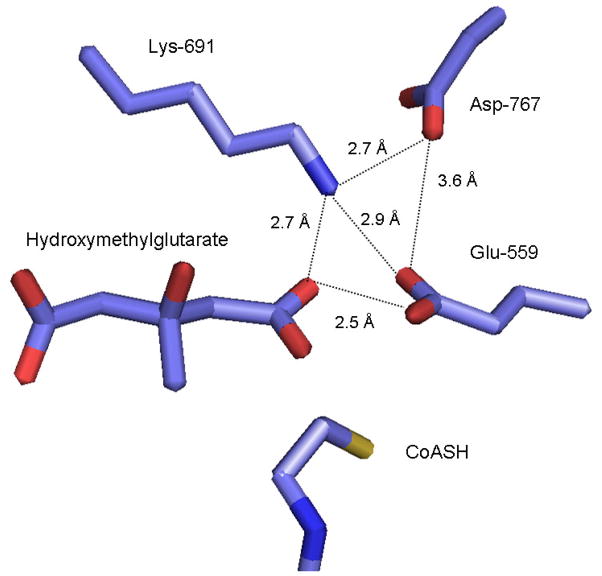
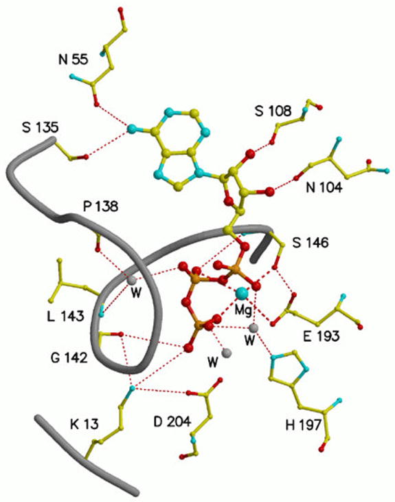
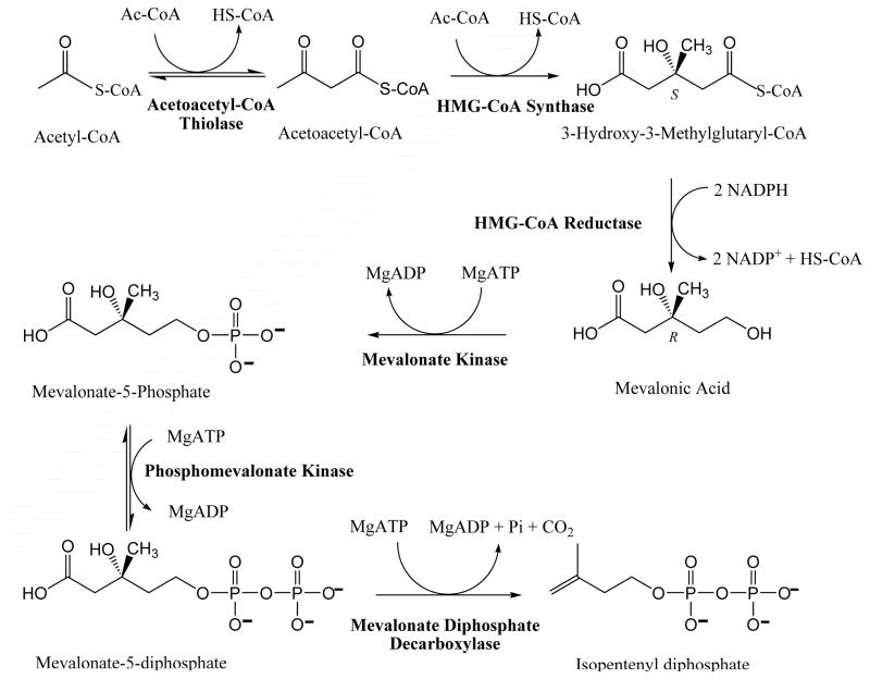
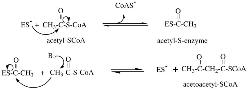
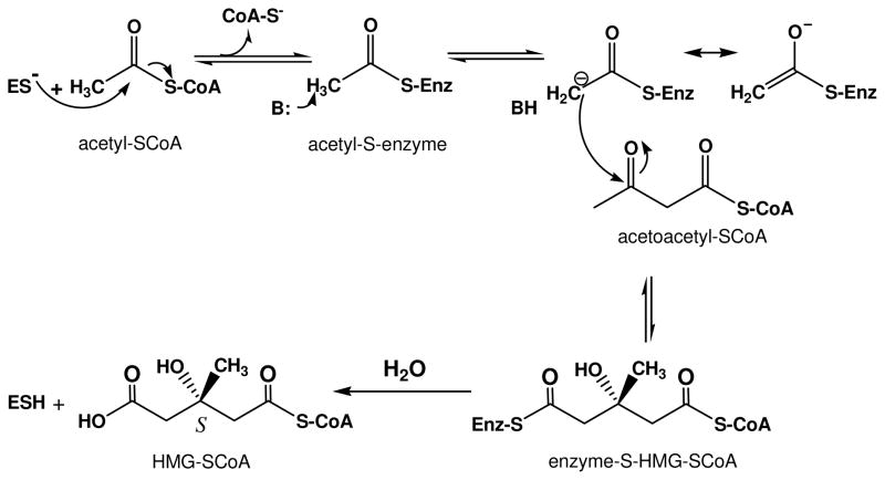
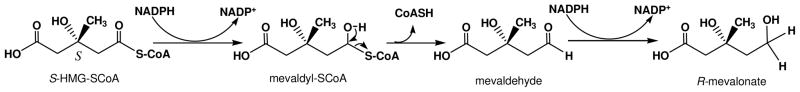
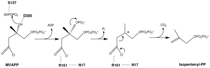
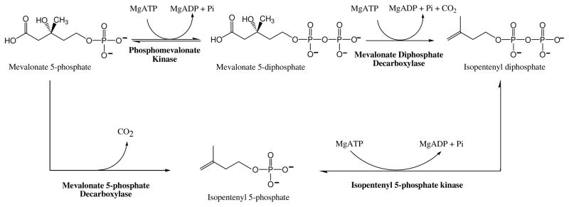
The mevalonate pathway accounts for conversion of acetyl-CoA to isopentenyl 5-diphosphate, the versatile precursor of polyisoprenoid metabolites and natural products. The pathway functions in most eukaryotes, archaea, and some eubacteria. Only recently has much of the functional and structural basis for this metabolism been reported. The biosynthetic acetoacetyl-CoA thiolase and HMG-CoA synthase reactions rely on key amino acids that are different but are situated in active sites that are similar throughout the family of initial condensation enzymes. Both bacterial and animal HMG-CoA reductases have been extensively studied and the contrasts between these proteins and their interactions with statin inhibitors defined. The conversion of mevalonic acid to isopentenyl 5-diphosphate involves three ATP-dependent phosphorylation reactions. While bacterial enzymes responsible for these three reactions share a common protein fold, animal enzymes differ in this respect as the recently reported structure of human phosphomevalonate kinase demonstrates. There are significant contrasts between observations on metabolite inhibition of mevalonate phosphorylation in bacteria and animals. The structural basis for these contrasts has also recently been reported. Alternatives to the phosphomevalonate kinase and mevalonate diphosphate decarboxylase reactions may exist in archaea. Thus, new details regarding isopentenyl diphosphate synthesis from acetyl-CoA continue to emerge.


Figures

Figure 1
Figure 1
Active site residue triad in Z. ramigera acetoacetyl-CoA thiolase, based on the structural coordinates 1DM3. The structure [14] indicates the acetyl-enzyme intermediate formed at Cys-89 as well as the acetyl-CoA that condenses with the reaction intermediate. His-348 interacts with the C1 carbonyl of bound acetyl–CoA to provide a charge sink that stabilizes the C2 carbanion formed after proton extraction by the general base Cis-378. The carbanion is in close proximity to C1 of the acetyl-enzyme intermediate and supports efficient condensation to form acetoacetyl-CoA and regenerate free enzyme.
Figure 2
Figure 2
Active site residue triad in S. aureus HMG-CoA synthase (mvaS), based on the structural coordinates 1XPL. The structure [18] depicts the acetyl-enzyme reaction intermediate formed at Cys-111/129 (bacterial/animal protein numbering). The enzyme’s second substrate, acetoacetyl-CoA is bound with His-233/264 interacting with C1 and C3 oxygens, providing a charge sink to facilitate attack by a carbanion on C3. This carbanion is produced upon abstraction of a proton from C2 of acetyl-enzyme by the general base Glu-79/95.
Figure 3
Figure 3
Active site residues in the soluble catalytic domain of human HMG-CoA reductase, based on the structural coordinates 1DQA. The structure [49] includes liganded hydroxymethylglutarate and indicates the positions of three residues (Asp-767, Lys-691, and Glu-559) implicated in enzyme function. Both lysine and glutamate would be in close proximity to the thioester carbonyl of HMG-CoA that is subjected to a two reductive steps, resulting in formation of the C5 alcohol of mevalonate. There are different proposals [48, 49] regarding the precise roles of these residues in substrate carbonyl polarization and/or the proton transfers that accompany NADPH reduction.
Figure 4
Figure 4
The MgATP binding site in rat mevalonate kinase [67], based on the structural coordinates 1KVK. ATP is bound in an anti conformation. Bound magnesium is coordinated to Glu-193 and Ser-146 as well as the beta and gamma phosphoryls of ATP. The catalytic residue Asp-204 is positioned to support transfer of the ATP gamma phosphoryl to an acceptor substrate. Conserved Lys-13 interacts with both Asp-204 and the gamma phosphoryl of substrate ATP. Dashed lines indicate coordination to magnesium; dotted lines indicate hydrogen bonds.
Figure 5
Figure 5
Feedback inhibitor binding to rat mevalonate kinase, based on the structural coordinates 2R42. Residue numbering is based on human mevalonate kinase. Inhibitor ligand electron density is fit using farnesyl thiodiphosphate (FSPP), although due to disorder or hydrolysis the beta phosphoryl is not well defined. The inhibitor binds in the ATP site [69] with its alpha phosphoryl group situated where the beta phosphoryl group of ATP would be located. Asp-204 and Ser-146 function as ligands to cation. Lys-13 would be expected to interact with the beta phosphoryl of inhibitor. The last 10 carbons of the farnesyl moiety interact with a number of nonpolar residues (e.g. Leu-53, Val-56, Val-133, Ile-196).
Figure 6
Figure 6
A solvent accessible surface representation of the feedback inhibitor (farnesylSP/farnesylSPP) binding site of mammalian mevalonate kinase, based on the structural coordinates 2R42. The animal MVK embeds farnesyl atoms C6-15 in a pocket to which the side chains of I196, T104, L53, N54, and I56 contribute [69]. This observation explains the high affinity (10−8 M; [56]) observed for feedback inhibition, which is competitive with respect to ATP. The image is adapted from Fu et al., Biochemistry 47 (2008) 3715–3724 [69], with permission of the American Chemical Society.
Figure 7
Figure 7
The active site cavity of human phosphomevalonate kinase and the location of conserved amino acid side chains implicated in enzyme function. The figure is based on the structural coordinates 3CH4, as reported by Chang et al. [85]. While the unliganded enzyme exhibits an open active site, the bound sulfate is interpreted as a phosphoryl group marker for the binding site of phosphorylated substrates. Sulfate is located in proximity to the N-terminal P-loop and interacts with the side chain of Arg-141, which has been shown [78] to influence ATP binding. Mutagenesis of Arg-18, Lys-22, Arg-84, Arg-110, and Arg-111 has resulted in observation of significant catalytic or substrate binding effects for these conserved residues.
Figure 8
Figure 8
A model of the ternary complex of human mevalonate diphosphate decarboxylase with ATP and mevalonate 5-diphosphate. A binary complex model was generated using the Z-dock algorithm (http://zdock.bu.edu) and coordinates of mevalonate diphosphate (from 2OI2) and the human enzyme (3D4J). The position of ATP is based on an overlay of a structure of the mevalonate kinase-ATP complex (1KVK) on the human MDD structure [95]. The predicted position of S127 agrees with the previous suggestion of its interaction with the phosphoryl chain of ATP [106]. The juxtapositioning of ATP’s gamma phosphoryl group with respect to MVAPP’s C3 oxygen is in accord with production of a 3-phosphoMVAPP reaction intermediate and supports the docking position of MVAPP in the binary complex model. The model predicts juxtapositioning Arg-161 and the C1 carboxyl of MVAPP. Asn-17 has been observed to hydrogen bond to Arg-161. The side chain of Asp-305 is close to the substrate’s C3 hydroxyl. Such interactions are in accord with functional roles for these conserved residues, as suggested by characterization of mutant MDD enzymes. The image has been adapted, with permission (Elsevier) from Voynova et al. Arch Biochem Biophys 480 (2008) 58–67.
Scheme 1
Scheme 1
Mevalonate (MVA) Pathway for Isopentenyl Diphosphate Biosynthesis.
Scheme 2
Scheme 2
Chemical steps in the biosynthetic acetoacetyl-CoA thiolase reaction.
Scheme 3
Scheme 3
Chemical steps in biosynthesis of HMG-CoA.
Scheme 4
Scheme 4
Chemical steps in the HMG-CoA reductase reaction.
Scheme 5
Scheme 5
Mevalonate diphosphate decarboxylase reaction chemistry and possible contributions of active site residues.
Scheme 6
Scheme 6
Proposed alternative reactions in biosynthesis of isopentenyl diphosphate.

References

    1. Bloch K. Science. 1965;150:19–28. - PubMed
    1. Eisenreich W, Bacher A, Arigoni D, Rohdich F. Cell Mol Life Sci. 2004;61:1401–1426. - PMC - PubMed
    1. Wilding EI, Brown JR, Bryant AP, Chalker AF, Holmes DJ, Ingraham KA, Iordanescu S, So CY, Rosenberg M, Gwynn MN. J Bacteriol. 2000;182:4319–4327. - PMC - PubMed
    1. Bochar FJDA, Stauffacher CV, Rodwell VW. Biosynthesis of mevalonic acid from acetyl-CoA. Pergamon Press; New York: 1999.
    1. Hedl M, Tabernero L, Stauffacher CV, Rodwell VW. J Bacteriol. 2004;186:1927–1932. - PMC - PubMed

Publication types

LinkOut - more resources