Review
. 2011 Jan-Feb;63(1-2):119-28.
doi: 10.1016/j.addr.2010.09.009. Epub 2010 Oct 7.

Intravital microscopy as a tool to study drug delivery in preclinical studies

Affiliations
Review

Intravital microscopy as a tool to study drug delivery in preclinical studies

Panomwat Amornphimoltham et al. Adv Drug Deliv Rev. 2011 Jan-Feb.

Abstract

The technical developments in the field of non-linear microscopy have made intravital microscopy one of the most successful techniques for studying physiological and pathological processes in live animals. Intravital microscopy has been utilized to address many biological questions in basic research and is now a fundamental tool for preclinical studies, with an enormous potential for clinical applications. The ability to dynamically image cellular and subcellular structures combined with the possibility to perform longitudinal studies have empowered investigators to use this discipline to study the mechanisms of action of therapeutic agents and assess the efficacy on their targets in vivo. The goal of this review is to provide a general overview of the recent advances in intravital microscopy and to discuss some of its applications in preclinical studies.


Figures

Figure 1
Figure 1. Multiphoton microscopy and second harmonic generation in live animals
A - Comparison between confocal and multiphoton microscopy. The salivary glands of a transgenic mouse expressing soluble GFP and a membrane-targeted peptide fused with the tandem-Tomato protein were imaged by using single-photon confocal microscopy (left panel, excitation wavelengths of 488 nm and 561 nm) or MPM (right panel, 930 nm excitation wavelength). Volume rendering of a z-stack of the first 60 µm from the surface of the glands (60x water immersion objective-Olympus). Bar 10 µm. B, C - Imaging the vasculature in live animals. 70 kDa Texas red was injected into the tail artery of anesthetized rats and then the salivary glands (B) or the brain cortex (C) were exposed and imaged by two-photon microscopy (excitation wavelength of 930 nm, 25X water immersion objective - Olympus). Volume rendering of a z-stack of the first 100 µm (B) and 700 µm (C) from the surface of the organ. Scale bars 10 µm (B) and 100 µm (C). D-Simultaneous excitation of multiple fluorophores by MPM. Salivary glands of anesthetized mice were exposed and Alexa 488 dextran and 594 transferrin were injected in the stroma. The glands were imaged by MPM (excitation wavelength of 740 nm, 60x water immersion objective-Olympus). Dextran (green) and transferrin (red) are accumulated in the stroma and internalized by stromal cells. In blue the endogenous fluorescence excited by MPM reveals the architecture of the acinar cells. Bar 50 µm. E MPM and SHG imaging of the salivary glands. The salivary glands of anesthetized mice were imaged to reveal the parenchyma of the glands (green) or the second harmonic signal generated by the collagen fibers (red) on the surface of the gland (excitation wavelengths of 720 nm and 800 nm, 20x water immersion objective Zeiss). Maximal projections of a z-stack from the first 100 µm below the surface are shown. F Endogenous fluorescence in live animals. Rat salivary glands were exposed and imaged by MPM (excitation wavelength of 740 nm, 60x water immersion objective-Olympus). A series of large intercalated duct are shown. Scale bar 40 µm.
Figure 2
Figure 2. Strategies for labeling molecules and the target issue for IVM
A QDs internalization in stromal cells. Transferrin conjugated with QDs (625 nm emission) were applied topically on the exposed salivary glands and after 20 minutes imaged by MPM (excitation wavelength 930 nm, 60x water immersion objective-Olympus).Transferrin is in red and collagen fibers in cyan. Bar 40 µm. B -Transgenic mice ubiquitously expressing soluble GFP (left), soluble Ds-Red (center) and tm-Tomato fused with a membrane targeted peptide (right) were imaged using confocal microscopy (left, 488 nm and right 561 nm excitation) or MPM (excitation wavelength of 930 nm). The left panel shows the submandibular salivary glands, the center panel tendon cells, and the right panel the liver. Bars 40 µm. C Gene delivery in rat salivary glands. Plasmid DNA encoding for mCherry TGN38, a marker for the trans-Golgi network (left panel, red) or GFP-actin (right panel green) were infused into the submandibular salivary glands and imaged as described in [83]. Bars 10 µm. D Cells engineered to express fluorescent proteins. Squamous cell carcinomas cells (OSCC3) were engineered to express the nuclear marker H2B fused with GFP (left panel), soluble mCherry, or the small GTPase Rab25 fused with GFP (right panel). Cells were orhtotopically implanted in the tongue of immunocompromised mice and imaged by MPM after 30 days (excitation wavelength of 930 nm). Left panel shows x-z maximum intensity projection from the first 70 µm below the surface of the tongue OSCC3 cells (green), stromal cells labeled by injection of Texas red dextran (red), and collagen fibers are shown in cyan. Right panel shows OSSC3-mCherry (red) and OSSC3-GFP-Rab25 (green) from a single plane at 100 µm below the surface of the tongue. Bars 10 and 30 µm.
Figure 3
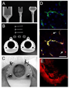
Figure 3. Devices for facilitating intravital microscopy in live animals
A- Custom-made holders for the stabilization of the externalized organs during IVM procedures (left and center holders are designed for the salivary glands, the right for the kidney). B Frames for the dorsal skin chamber made of the biocompatible polyacetal resin Duracon [130]. C- Dorsal skin chamber implanted in the back of a nude mouse. The vasculature is clearly visible. D Imaging the vasculature in the salivary glands of live rats. The salivary glands of an anesthetized rat were exposed, placed in a custom-made holder, and imaged by MPM as described in [111]. First, Hoechst (blue) and 500 kDa FITC-dextran (green) were administered systemically (upper panel) followed by a second injection of 70 kDa Texas Red-dextran (red). Note that after 20 minutes from the second injection, the larger dextran is retained in the vasculature, whereas the smaller one diffuses into the stroma and is internalized by stromal cells (arrows) (excitation wavelength of 820 nm, 60x water immersion objective-Olympus). Bar 30 µm. E The tumor vasculature imaged through the dorsal skin chamber. OSCC3 cells were implanted in the dorsal skin chamber mounted on the back of a nude mouse. 70 kDa Texas Red dextran was injected in the tail vein of the mouse and imaged by MPM (excitation wavelength of 930 nm, 20X water immersion objective from Olympus).

References

    1. Hoffman RM, Yang M. Whole-body imaging with fluorescent proteins. Nat Protoc. 2006;1:1429–1438. - PubMed
    1. Hyafil F, Cornily JC, Feig JE, Gordon R, Vucic E, Amirbekian V, Fisher EA, Fuster V, Feldman LJ, Fayad ZA. Noninvasive detection of macrophages using a nanoparticulate contrast agent for computed tomography. Nat Med. 2007;13:636–641. - PubMed
    1. Mitra A, Nan A, Line BR, Ghandehari H. Nanocarriers for nuclear imaging and radiotherapy of cancer. Curr Pharm Des. 2006;12:4729–4749. - PubMed
    1. Mulder WJ, Strijkers GJ, van Tilborg GA, Griffioen AW, Nicolay K. Lipid-based nanoparticles for contrast-enhanced MRI and molecular imaging. NMR Biomed. 2006;19:142–164. - PubMed
    1. Yang M, Jiang P, Hoffman RM. Whole-body subcellular multicolor imaging of tumor-host interaction and drug response in real time. Cancer Res. 2007;67:5195–5200. - PubMed

Publication types