. 2011 Feb;41(2):318-28.
doi: 10.1016/j.nbd.2010.09.020. Epub 2010 Oct 13.

Modification of ubiquitin-C-terminal hydrolase-L1 by cyclopentenone prostaglandins exacerbates hypoxic injury

Affiliations

Modification of ubiquitin-C-terminal hydrolase-L1 by cyclopentenone prostaglandins exacerbates hypoxic injury

Hao Liu et al. Neurobiol Dis. 2011 Feb.

Abstract

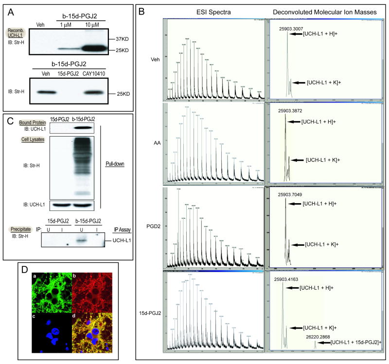
Cyclopentenone prostaglandins (CyPGs), such as 15-deoxy-Δ(12,14) -prostaglandin J(2) (15d-PGJ(2)), are active prostaglandin metabolites exerting a variety of biological effects that may be important in the pathogenesis of neurological diseases. Ubiquitin-C-terminal hydrolase L1 (UCH-L1) is a brain specific deubiquitinating enzyme whose aberrant function has been linked to neurodegenerative disorders. We report that [15d-PGJ(2)] detected by quadrapole mass spectrometry (MS) increases in rat brain after temporary focal ischemia, and that treatment with 15d-PGJ(2) induces accumulation of ubiquitinated proteins and exacerbates cell death in normoxic and hypoxic primary neurons. 15d-PGJ(2) covalently modifies UCH-L1 and inhibits its hydrolase activity. Pharmacologic inhibition of UCH-L1 exacerbates hypoxic neuronal death while transduction with a TAT-UCH-L1 fusion protein protects neurons from hypoxia. These studies indicate that UCH-L1 function is important in hypoxic neuronal death and that excessive production of CyPGs after stroke may exacerbate ischemic injury by modification and inhibition of UCH-L1.


Conflict of interest statement

Disclosure/Conflict of Interest: The authors declare no conflict of interest. Contents do not represent the views of the Department of Veterans Affairs or the United States Government.

Figures

Figure 1
Figure 1. [15-deoxy-Δ12,14-PGJ2(15d-PGJ2)] in rat brain after middle cerebral artery occlusion (MCAO)
A. MS/MS fragmentation pattern of 15d-PGJ2 authentic standard with a Parent Mass (m/z) of 316. The prevalent 271m/z daughter Ion was used to quantify 15d-PGJ2 using quadrupole MS/MS with selective reaction monitoring. Arrow (inset) indicates fragmentation point of parent ion. B. 15d-PGJ2 concentration in rat brain hemisphere 24h after 90 minutes MCAO. Data are expressed as means +/- SE. *P < .05. n=4-6.
Figure 2
Figure 2. Effect of treatment with 15d-PGJ2 upon accumulation of Ub-proteins and cell death in primary neuronal culture
A. left: Primary neurons were incubated with 15d-PGJ2 for 24h then harvested. Ub-proteins were detected by IB with anti-ubiquitin antibody. β-actin served as a loading control. right: representative confocal images (240×) of primary neurons after incubation with vehicle (veh) or 20 μM 15d-PGJ2 for 24h prior to staining with anti-ubiquitinated protein antibody paired with FITC-conjugated secondary antibody (green) and DAPI (blue). B. Primary neurons were incubated with vehicle or 15d-PGJ2 for 1 h prior to normoxia (a,b) or hypoxia (c,d). Cell death was measured using LDH (a,c) and MTT (b,c) methods. n = 6 wells per group. * P < 0.02. C. left: Primary neurons were incubated with vehicle, CAY10410 or 15d-PGD2 for 24h prior to harvest. Ub-proteins were detected by IB with anti-ubiquitin antibody. n= 6 per group. right: Primary neurons were treated with CAY10410 or 15d-PGD2 1 h prior to hypoxia. Cell death was measured using the LDH method. D: Primary neurons were incubated with vehicle, 15d-PGJ2 or CAY10410 (20 μM) for 24h prior to hypoxia or normoxia treatment. 24 h later, cells were stained with propidium iodide (PI) and Hoechst. a: PI-positive cells were counted and cell death was calculated as percent total cells (Hoechst). n = 10 – 12 per group. b: representative photos of Hoechst and PI-Hoechst overlay stained cells. Single arrows indicate representative cells with apoptotic morphology; double arrows are representative cells exhibiting rounded morphology. c: Percentage cells with normal morphology in normoxic (top) and hypoxic (bottom) groups treated with 20 μM of 15d-PGJ2, 15d-PGD2, CAY10410 or vehicle. * P < 0.001; ** < 0.01; # < .05; NS: not significant. Data are expressed as means +/- SE. IB: immunoblot; MK: 1 μM MK801; SP: 20 μM staurosporin; Ub-Proteins: Ubiquitinated proteins; Veh: vehicle.
Figure 3
Figure 3. Covalent modification of UCH-L1 by 15d-PGJ2 in vitro and in primary neurons
A. upper: Recombinant UCH-L1 protein (1 μg) was incubated with biotinylated (b-) 15d-PGJ2 or vehicle (ethanol, Veh) for 2h. b-15d-PGJ2-UCH-L1 adducts were detected by immunoblotting with streptavidin-HRP (Str-H). lower: Recombinant UCH-L1 protein (1 μg) was preincubated with methyl acetate (Veh), 500 μM 15d-PGJ2 or CAY10410 for 30 min before incubation with 5 μM b-15d-PGJ2 and b-15d-PGJ2-UCH-L1 adduct in each reaction were detected. B. Capillary LC ESI-TOF MS analysis of UCH-L1 incubated with Vehicle (Veh), arachidonic acid (AA), PGD2 and 15d-PGJ2. Charge state distribution (left) and deconvoluted molecular ion mass spectra (right) are shown. C. Primary neurons were incubated with 10 μM 15d-PGJ2 or b-15d-PGJ2 for 2h before harvest. The b-15d-PGJ2-UCH-L1 adducts in cell lysates were detected after Avidin pull-down or immunoprecipitation assays. Top: Avidin pull-down assay: Proteins bound to NeutrAvidin beads were separated by SDS-PAGE and blotted with anti-UCH-L1 antibody. Center: Immunoblots of cell lysates probed with Streptavidin-HRP (Str-H) and anti-UCH-L1 antibody. Bottom: IP assay: Primary neuron cell lysates were immunoprecipitated with anti-UCH-L1 antibody (U) or control IgG (I) and protein A/G beads. Immunoprecipitates were separated and probed with Streptavidin-HRP to detect b-15d-PGJ2-UCH-L1 adducts. D. Immunocytochemical staining of primary neurons after 2h incubation with 10μM b-15d-PGJ2. Representative photos of: a) endogenous UCHL-1 distribution using FITC conjugated anti-UCHL-1 antibody. b) b-15d-PGJ2 distribution using Cy3 conjugated anti-biotin antibody. c) DAPI staining of nuclei. d) Merged picture. Photos at 60×. IB: Immunoblot; IP: immunoprecipitation.
Figure 4
Figure 4. Effect of 15d-PGJ2 on UCH-L1 hydrolase activity
1μg of recombinant UCH-L1 was incubated with 15d-PGJ2, CAY10410 or vehicle (Veh, methyl acetate) at indicated concentrations. Hydrolase activity was measured using the fluorescent substrate ubiquitin-AMC. Fluorescence intensity (arbitrary units, λex=360nm and λem=460nm) produced by cleavage of ubiquitin-AMC was recorded at indicated time points. Mutant UCH-L1 C90S lacking hydrolase activity was included as a negative control. Data shown is the mean of the experiment with two replicates. Veh: vehicle; C90S: recombinant UCH-L1 C90S protein.
Figure 5
Figure 5. Effect of UCH-L1 activity on hypoxic neuronal cell death and Ub-protein accumulation in primary neurons
A. Primary neurons were treated with vehicle (DMSO), LDN 57444 or untreated for 1 hour, then underwent normoxia (left) or 3h hypoxia (right) then returned to normoxic conditions for 24h. Cell death was measured by LDH assay. N= 6 wells per group. Data are expressed as means +/- SE. * P < 0.01; ** < 0.05 vs vehicle treated group. B. Representative immunoblots of primary neuronal cell lysates (treated as in A) probed with anti-ubiquitin and anti-actin (loading control) antibodies. right: graphic illustration of western blot ubiquitinated protein lane densities expressed as % vehicle. n=3. * P < 0.05. C. top: Structure diagrams of HA-tagged wild type TAT-UCH-L1 WT and mutant TAT-UCH-L1 C90S fusion proteins. bottom left: TAT-UCH-L1 WT or mutant TAT-UCH-L1 C90S fusion proteins detected with immunoblots probed with anti-UCH-L1 antibody after incubation in primary neurons. (a-c, bottom right): Representative photos of immunocytochemical staining of fusion protein transduction in primary neurons after 1h incubation with 0.08μM of recombinant TAT-UCH-L1 WT, mutant TAT-UCH-L1 C90S proteins or PBS (UN) using anti-HA antibody (green) and DAPI nuclear stain (blue). D: Primary neurons were incubated with TAT-UCH-L1 WT, TAT-UCH-L1 C90S fusion proteins or vehicle for 1h prior to hypoxia. After hypoxia, cells were returned to normoxic conditions for 24h. Cell death was measured by assaying LDH released into the medium. N=6 per group. Data are expressed as means ± SE. * P < 0.01 versus vehicle-treated group. LDN: UCH-L1 inhibitor LDN 57444; Veh: vehicle; IB: immunoblot; WT: wild type TAT-UCH-L1; C90S: mutant TAT-UCH-L1 C90S; MK: MK801 1 μM; SP: staurosporin 20 μM; UN: untreated.
Figure 6
Figure 6. Modification of UCH-L1 and other proteins by arachidonic acid, prostaglandins and their metabolites in primary neurons
A. Primary neurons were incubated with biotinylated arachidonic acid (AA), prostaglandins (PGD2 and PGE2), or CyPGs (15d-PGJ2 and PGA1) at indicated concentrations for 2h under normal culture conditions. Modified protein in the cell lysates were detected by streptavidin-HRP. B. Avidin pull-down assay detecting AA metabolite-modified UCH-L1 in primary neurons. Neurons were incubated with 20μM biotinylated AA and subjected to hypoxia (+) or normoxia (-) before harvest at the indicated time points. Top: NeutrAvidin bead bound UCH-L1 was detected by IB with anti-UCH-L1 antibody. Bottom: Biotinylated proteins and endogenous UCHL-L1 in neuronal lysates were detected by IB with streptavidin-HRP and anti-UCH-L1 antibody respectively. C. The effect of COX-2 overexpression on AA metabolite-modified protein accumulation in primary neurons. Neurons infected with AdV-COX-2, AdV-EGFP or vehicle (Veh) were incubated with 20μM biotinylated AA and subjected to hypoxia or normoxia. 3h after hypoxia/normoxia, cells were harvested and biotinylated proteins were detected by IB with streptavidin-HRP. D. The effect of COX-2 inhibition on AA metabolite-modified protein accumulation in primary neurons. Neurons were treated with rofecoxib (1μM) or vehicle (-) for 1h prior to incubation with 20μM biotinylated AA. Cells were harvested after hypoxia (+) or normoxia (-). Biotinylated proteins were detected by IB with streptavidin-HRP. E. Avidin pull-down assay to detect PGD2 metabolite-modified UCH-L1 in primary neurons. Neurons were incubated with 10μM biotinylated PGD2 and subjected to hypoxia (+) or normoxia (-). Cells were harvested 0 and 24h after hypoxia/normoxia. Upper three panels: Biotinylated proteins and endogenous UCH-L1 proteins in neuronal lysates were detected by IB with streptavidin-HRP (arrow at band indicating MW of UCH-L1) and anti-UCH-L1 antibody respectively. Bottom panel: NeutrAvidin bead-bound UCH-L1 was detected by IB with anti-UCH-L1 antibody. M: marker; AA: Arachidonic acid; UN: untreated cells; IB: immunoblot; AdV-COX-2: adenovirus containing mouse WT COX-2; AdV-EGFP: adenovirus containing EGFP.

Similar articles

Cited by

References

    1. Ahmad M, Zhang Y, Liu H, Rose ME, Graham SH. Prolonged opportunity for neuroprotection in experimental stroke with selective blockade of cyclooxygenase-2 activity. Brain Res. 2009;1279:168–73. - PMC - PubMed
    1. Aldini G, Carini M, Vistoli G, Shibata T, Kusano Y, Gamberoni L, Dalle-Donne I, Milzani A, Uchida K. Identification of actin as a 15-deoxy-Delta12,14-prostaglandin J2 target in neuroblastoma cells: mass spectrometric, computational, and functional approaches to investigate the effect on cytoskeletal derangement. Biochemistry. 2007;46:2707–18. - PubMed
    1. Cao G, Pei W, Ge H, Liang Q, Luo Y, Sharp FR, Lu A, Ran R, Graham SH, Chen J. In Vivo Delivery of a Bcl-xL Fusion Protein Containing the TAT Protein Transduction Domain Protects against Ischemic Brain Injury and Neuronal Apoptosis. J Neurosci. 2002;22:5423–31. - PMC - PubMed
    1. Choi J, Levey AI, Weintraub ST, Rees HD, Gearing M, Chin LS, Li L. Oxidative modifications and down-regulation of ubiquitin carboxyl-terminal hydrolase L1 associated with idiopathic Parkinson's and Alzheimer's diseases. J Biol Chem. 2004;279:13256–64. - PubMed
    1. Colgan L, Liu H, Huang SY, Liu YJ. Dileucine motif is sufficient for internalization and synaptic vesicle targeting of vesicular acetylcholine transporter. Traffic. 2007;8:512–22. - PubMed

Publication types

MeSH terms