. 2011 Jan;32(1):39-47.
doi: 10.1016/j.biomaterials.2010.08.103. Epub 2010 Oct 12.

The spreading, migration and proliferation of mouse mesenchymal stem cells cultured inside hyaluronic acid hydrogels

Affiliations

The spreading, migration and proliferation of mouse mesenchymal stem cells cultured inside hyaluronic acid hydrogels

Yuguo Lei et al. Biomaterials. 2011 Jan.

Abstract

Synthetic hydrogel scaffolds that can be used as culture systems that mimic the natural stem cell niche are of increased importance for stem cell biology and regenerative medicine. These artificial niches can be utilized to control the stem cell fate and will have potential applications for expanding/differentiating stem cells in vitro, delivering stem cells in vivo, as well as making tissue constructs. In this study, we synthesized hyaluronic acid (HA) hydrogels that could be degraded through a combination of cell-released enzymes and used them to culture mouse mesenchymal stem cells (mMSC). To form the hydrogels, HA was modified to contain acrylate groups and crosslinked through Michael addition chemistry using non-degradable, plasmin degradable or matrix metalloproteinase (MMP) degradable crosslinkers. Using this hydrogel we found that mMSC proliferation occurred in the absence of cell spreading, that mMSCs could only spread when both RGD and MMP degradation sites were present in the hydrogel and that mMSCs in hydrogels with high density of RGD (1000 μm) spread and migrated faster and more extensively than in hydrogels with low density of RGD (100 μm).


Figures

Figure 1
Figure 1
The storage (G') and loss modulus (G") of HA-MMP hydrogels. (A) 3% HA hydrogels with various crosslinking densities (r = 0.25, 0.4, 0.5). (B) 3%, 4%, 5% HA hydrogels at r = 0.25. The modulus were measured with a rheometer at constant strain of 0.05 with frequency ranging from 0.1 to 10 rad/s. The average of G' and G" over the frequency range were shown in (C)
Figure 2
Figure 2
Effect of crosslinker identity on D1 cells in HA hydrogels. (A–F) Cell viability at day 3 (green: live cell; red: dead cell). (G–L) Cell migration from fibrin/cell clot into hydrogels. Snapshots were taken at day 3. The dash lines highlight the edges of the clots. “C” refers to fibrin/cell clot. (M) Proliferation rates of cells in hydrogels with 100 µM RGD. Crosslinker: DTT for (A, B, G, H); HS-Plasmin-SH for (C, D, I, J); HS-MMP-SH for (E, F, K, L). RGD concentration: 100 µM for (A, C, E, G, I, K); 1000 µM for (B, D, F, H, J, L). Scale bar: 100 µm. 3.5% HA and r = 0.4 for all gels
Figure 3
Figure 3
Effect of RGD concentration on D1 cells in HA-MMP hydrogels. (A) Cell viability and spreading at day 3 (green: live cell; red: dead cell). (B) Proliferation rates. (C) Cell migration at day 5. The dash lines highlight the edges of the clots. “C” refers to fibrin/cell clot. (D) Migration rates were 0, 55, 68, 77, 72 and 87 µm/day for cells in the gels with 0, 100, 250, 500, 750 and 1000 µM RGD respectively. Scale bars for (A) and (C) are 200 and 100 µm, respectively. 3% HA and r = 0.4 for all gels
Figure 4
Figure 4
Effect of HA concentration on D1 cells in HA-MMP hydrogels. (A) Cell viability and spreading at day 2 (green: live cell; red: dead cell). (B) Proliferation rates of cells in 3%, 4% and 5% HA gels with 100 µM RGD. (C) Cell migration. Snapshots were taken at day 6. The dash lines highlight the edges of the clots. “C” refers to the fibrin/cell clot (D) Migration rates were 95, 50, 0 µm/day in 3%, 4%, 5% hydrogels with 100 µM RGD (open symbols) and were 99, 88, 2 µm/day in 3%, 4%, 5% hydrogels with 1000 µM RGD (filled symbols), respectively. Scale bars for (A) and (C) are 200 and 100 µm, respectively. r = 0.25 for all gels.
Figure 5
Figure 5
Effect of crosslinking density on D1 cells inside HA-MMP hydrogels. (A) Cell viability and spreading at day 2 (green: live cell; red: dead cell). (B) Proliferation rates of cells in hydrogels with 100 µM RGD. (C) Cell migration. Snapshots were taken at day 3. The dash lines highlight the edges of the clots. “C” refers to the fibrin/cell clot (D) Migration rates were 220, 220, 210 µm/day in hydrogels at r = 0.25, 0.4, 0.5 at 100 µM RGD (open symbols) and were 240, 160, 160 µm/day hydrogels at r = 0.25, 0.4, 0.5 at 1000 µM RGD (filled symbols), respectively. Scale bars for (A) and (C) are 200 and 100 µm, respectively. 3% HA for all gels
Scheme 1
Scheme 1
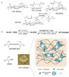
(A) Synthesis of HA-AC and (B) Making HA hydrogels through Michael Addition

References

    1. Weissman IL. Stem cells: units of development, units of regeneration, and units in evolution. Cell. 2000;100(1):157–168. - PubMed
    1. Passier R, van Laake LW, Mummery CL. Stem-cell-based therapy and lessons from the heart. Nature. 2008;453(7193):322–329. - PubMed
    1. Kim SU, de Vellis J. Stem cell-based cell therapy in neurological diseases: a review. Neurosci . Res . 2009;87(10):2183–2200. - PubMed
    1. Thomson JA, Itskovitz-Eldor J, Shapiro SS, Waknitz MA, Swiergiel JJ, Marshall VS, et al. Embryonic stem cell lines derived from human blastocysts. Science. 1998;282(5391):1145–1147. - PubMed
    1. Reubinoff BE, Pera MF, Fong CY, Trounson A, Bongso A. Embryonic stem cell lines from human blastocysts: somatic differentiation in vitro. Nat . Biotechnol . 2000;18(4):399–404. - PubMed