. 2010 Dec 17:1366:233-45.
doi: 10.1016/j.brainres.2010.10.009. Epub 2010 Oct 8.

Sex differences in β-amyloid accumulation in 3xTg-AD mice: role of neonatal sex steroid hormone exposure

Affiliations

Sex differences in β-amyloid accumulation in 3xTg-AD mice: role of neonatal sex steroid hormone exposure

Jenna C Carroll et al. Brain Res. .

Abstract

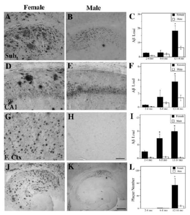
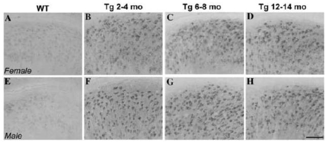
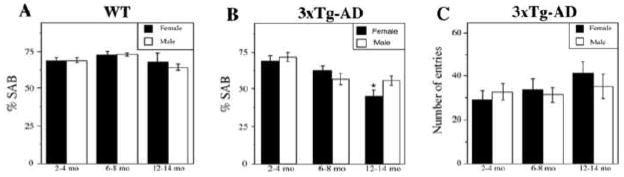
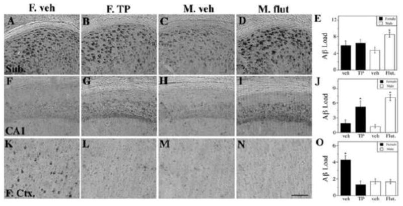
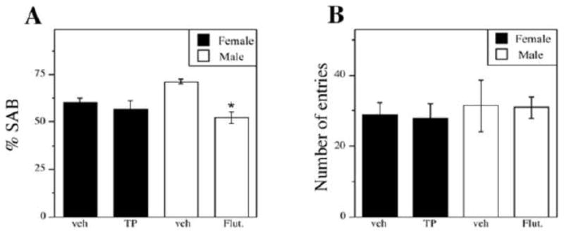
The risk of Alzheimer's disease (AD) is higher in women than in men, a sex difference that likely results from the effects of sex steroid hormones. To investigate this relationship, we first compared progression of β-amyloid (Aβ) pathology in male and female triple transgenic (3xTg-AD) mice. We found that female 3xTg-AD mice exhibit significantly greater Aβ burden and larger behavioral deficits than age-matched males. Next, we evaluated how the organizational effects of sex steroid hormones during postnatal development may affect adult vulnerability to Aβ pathology. We observed that male 3xTg-AD mice demasculinized during early development exhibit significantly increased Aβ accumulation in adulthood. In contrast, female mice defeminized during early development exhibit a more male-like pattern of Aβ pathology in adulthood. Taken together, these results demonstrate significant sex differences in pathology in 3xTg-AD mice and suggest that these differences may be mediated by organizational actions of sex steroid hormones during development.


Figures

Figure 1
Figure 1
Female 3xTg-AD mice exhibit higher levels of Aβ than males. Representative images show Aβ immunoreactivity from 12–14 mo female (A, D, G) and male (B, E, H) 3xTg-AD mice in the subiculum (A,B), hippocampus CA1 (D,E), and frontal cortex (G,H). Scale bar = 100μm. Quantification of Aβ immunoreactive load shows significant differences between females (solid bars) and males (open bars) in the subiculum (C) [F (7,46) = 23.72, p < 0.0001], hippocampus CA1 (F) [F (7,46) = 5.88, p = 0.0005], and frontal cortex (I) [F (7,46) = 9.76, p <0.0001]. Representative low magnification images of Aβ immunoreactivity in hippocampus and subiculum of 12–14 mo female (J) and male (K) 3xTg-AD mice show significantly higher numbers of Aβ plaques in females (L) [F (8,43) = 9.25, p < 0.0001]. Scale bar = 250μm. Data show mean values ± SEM. * p < 0.01 relative to males of same age group.
Figure 2
Figure 2
No difference in levels of amyloid precursor protein (APP) C-terminal fragments (CTFs) were observed between male and female 3xTg-AD mice. Representative images show CTF immunoreactivity (CT20 antibody) in the subiculum of male and female non-transgenic C57Bl6/129S and 3xTg-AD mice. Non-transgenic mice (A–E) show low levels of CTF-immunoreactivity while both male (F–H) and female (B–D) mice show similar, high levels of CTF- immunoreactivity. Scale bar = 100μm.
Figure 3
Figure 3
Female 3xTg-AD mice perform more poorly than males on spontaneous alternation behavior (SAB), a hippocampal-dependent working memory task. (A) SAB performance did not decline with age or significantly differ by sex in non-transgenic C57Bl6/129S (WT) mice. (B) We observed an age-related decline in SAB performance in both females (solid bars) [F (3,20) = 0.87, p = 0.002] and males (open bars) [F (3, 7) = 0.67, p = 0.031]. This effect is significantly different between females and males in the 12–14 mo age group [F (3,19) = 6.58, p = 0.0001]. * p < 0.05 from males of same age group. (C) As a control for activity level, the number of arm entries was measured; it was not significantly different across groups [F (3,19) = 0.79, p = 0.56]. Data show mean values ± SEM.
Figure 4
Figure 4
Neonatal flutamide treatment demasculinizes male 3xTg-AD adult brains and TP treatment defeminizes female brains in terms of Aβ pathology in a region-specific manner. Representative photomicrographs display Aβ immunoreactivity visualized in the subiculum (AD), CA1 of hippocampus (F–I), and frontal cortex (K-N). Scale bar = 100μm. Significant treatment differences were observed in quantification of Aβ immunohistochemistry load in the subiculum (E) [F (4,32) = 2.82, p = 0.05], the CA1 (J) [F (4,32) = 17.11, p < 0.0001] and the frontal cortex (O) [F (4,32) = 13.01, p < 0.0001]. Data are represented as mean values ± SEM. * p < 0.05 from opposite sex of same age group.
Figure 5
Figure 5
Neonatal flutamide treatment, but not TP treatment alters hippocampal-dependent behavior on the Y-maze. Neonatally-hormone treated mice exhibited a significant overall treatment effect on Y-maze performance (A) [F (3,30) = 6.37, p = 0.003]. The number of arm entries was not significantly different across groups (B). Data are represented as mean values ± SEM. * p < 0.05 from opposite sex of same age group.

Similar articles

Cited by

References

    1. Akhmadeev AV, Kalimullina LB. [Neonatal androgenization of female rats during sex differentiation of the brain modifies neuronal organization of the amygdala complex] Ontogenez. 2005;36:64–7. - PubMed
    1. Akhondzadeh S, Rezaei F, Larijani B, Nejatisafa AA, Kashani L, Abbasi SH. Correlation between testosterone, gonadotropins and prolactin and severity of negative symptoms in male patients with chronic schizophrenia. Schizophr Res. 2006;84:405–10. - PubMed
    1. Alkayed NJ, Murphy SJ, Traystman RJ, Hurn PD, Miller VM. Neuroprotective effects of female gonadal steroids in reproductively senescent female rats. Stroke. 2000;31:161–8. - PubMed
    1. Anahara R, Toyama Y, Mori C. Flutamide induces ultrastructural changes in spermatids and the ectoplasmic specialization between the Sertoli cell and spermatids in mouse testes. Reprod Toxicol. 2004;18:589–96. - PubMed
    1. Andersen K, Launer LJ, Dewey ME, Letenneur L, Ott A, Copeland JRM, Dartigues J-F, Kragh-Sorensen P, Baldereschi M, Brayne C, Lobo A, Martinez-Lage JM, Stijnen T, Hofman A. Gender differences in the incidence of AD and vascular dementia: The EURODEM Studies. Neurology. 1999;53:1992–7. - PubMed

Publication types

MeSH terms