. 2011 Jan 7;405(1):3-17.
doi: 10.1016/j.jmb.2010.09.064. Epub 2010 Oct 8.

An RNA conformational switch regulates pre-18S rRNA cleavage

Affiliations

An RNA conformational switch regulates pre-18S rRNA cleavage

Allison C Lamanna et al. J Mol Biol. .

Abstract

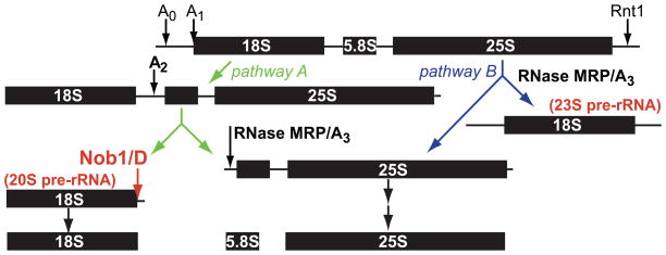
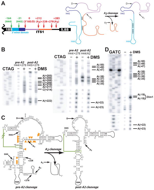
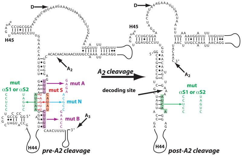
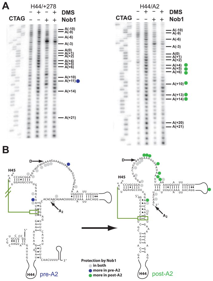
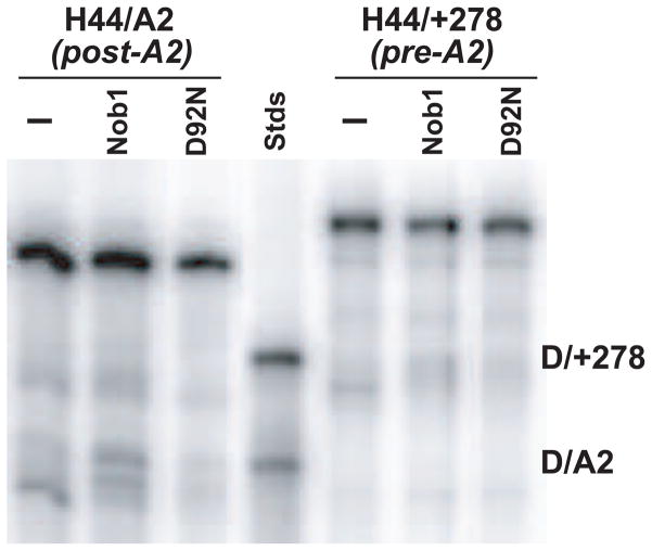
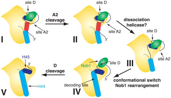
To produce mature ribosomal RNAs (rRNAs), polycistronic rRNA transcripts are cleaved in an ordered series of events. We have uncovered the molecular basis for the ordering of two essential cleavage steps at the 3'-end of 18S rRNA. Using in vitro and in vivo structure probing, RNA binding and cleavage experiments, and yeast genetics, we demonstrate that a conserved RNA sequence in the spacer region between the 18S and 5.8S rRNAs base-pairs with the decoding site of 18S rRNA in early assembly intermediates. Nucleolar cleavage at site A(2) excises this sequence element, leading to a conformational switch in pre-18S rRNA, by which the ribosomal decoding site is formed. This conformational switch positions the nuclease Nob1 for cytoplasmic cleavage at the 3'-end of 18S rRNA and is required for the final maturation step of 18S rRNA in vivo and in vitro. More generally, our data show that the intrinsic ability of RNA to form stable structural switches is exploited to order and regulate RNA-dependent biological processes.


Figures

Figure 1
Figure 1
Cartoon of pre-rRNA cleavage pathways. For simplicity 60S processing pathways are not indicated. Cleavage sites discussed herein are shown in bold. Pathway A shows cleavage at sites A0, A1, and A2 yielding 20S pre-rRNA, before cleavages at D and A3, resulting in production of mature 18S and 25S rRNA. Alternatively, cleavage at site A3 without prior cleavage at sites A0-A2 (pathway B) produces mature 25S rRNA and the 23S pre-rRNA, which is not converted into 18S rRNA.
Figure 2
Figure 2
pre-rRNAs prior to and after cleavage at site A2 are in different conformations. (A) Schematic showing pre-rRNA constructs used in this study, comprising the 3′ minor domain of 18S and the 5′ end of ITS1. Green arrows represent 5′-ends, and red arrows represent 3′-ends of constructs. Color-coded schematic of the RNA conformational change after A2-cleavage is included for clarity. (B) Examples of in vitro DMS probing data by primer extension. H44/+278 rRNAs are in the pre-A2 cleavage structure, while H44/A2 and H45/+278 rRNAs are in the post-A2 cleavage structure. Reverse transcriptions were carried out with oligos d (left) and b (right panel). Note that reverse transcriptase stops prior to the modified nucleotide while ddNTP incorporation leads to a stop after the incorporated nucleotide. Thus, the sequencing ladder is upshifted 1 nt relative to the DMS lane. Labeled residues show increased accessibility in the pre-A2 cleavage structure. (C) Mapping of the data in (B) onto secondary structure models for the two pre-rRNA conformers: pre-A2 cleavage (H44/+278) and post-A2 cleavage (H44/A2). Residues equally accessible to DMS are shown by gray circles, residues more accessible in the pre-A2 cleavage structure are shown in orange triangles. Open circles show mild accessibility as compared to filled circles. The olive line indicates a tertiary interaction observed in the 30S crystal structures. This interaction is expected to be disrupted in the pre-A2 structure, as indicated by the strike-through. Arrows represent the ends of in vitro transcribed RNAs used herein and shown in (A). All numbering is relative to site D. See Figs. S2 and S3A for full DMS probing gels and line scan quantitations. (D) The 23S pre-rRNA is in the pre-A2 cleavage conformation. In vivo DMS probing of 23S pre-rRNA. Yeast strain YKK37 has the bms1 gene under the control of a galactose-inducible promoter and was grown in YP-glucose overnight at 30 °C to deplete Bms1. Primer extension using oligo b is shown. Compare to pre-A2/+DMS lane in (B), right panel. The conserved, Dim1-methylated adenosines in H45 (−18/−19) are indicated. Native methylation of these residues in 20S rRNA leads to a strong reverse transcription stop even in the absence of DMS , which is not present in 23S, consistent with Dim1-dependent methylation occurring late in the cytoplasm . Lack of primer extension above the Dim1 site in 20S rRNA also means that all readings above that region arise from the 23S rRNA in the sample obtained in the absence of Bms1, even though there is substantial amount of 20S remaining in our sample (46%). Refer to Figure S5 for the full DMS gel and Northern analysis of the rRNA from these strains. Figure S3B has linescan quantitations.
Figure 3
Figure 3
pre-rRNA mutations used in this study. Highlighted residues are changed to the indicated sequence. Mut A and B destabilize the inhibitory structure. Mut S stabilizes the inhibitory structure. Mut αS1 and αS2 in combination with mut S remove the stabilization. Mut N have no effect on the inhibitory or mature structure, but mutate the same residues mutated in Mut S.
Figure 4
Figure 4
Nob1 interacts differently with pre-rRNAs prior to and after cleavage at site A2. (A) Examples of DMS probing data by primer extension with oligo b, as described in Figure 2. Blue circles indicate residues more accessible in the pre-A2 (than in the post-A2) structure, and green circles represent residues that are more accessible in the post-A2 (than in the pre-A2) structure. (B) Mapping of the data in (A) onto the secondary structure models for the two pre-rRNA conformers: H44/+278 (pre-A2) and H44/A2 (post-A2), as in Figure 2. See Fig. S2 for full DMS probing gels, and Figure S6 for linescan quantitations.
Figure 5
Figure 5
Nob1 only cleaves RNA in the post-A2 structure. 0.2 μM Nob1, D92N/Nob1, or no protein was incubated with 3′-end labeled H44/A2 (post-A2 structure) or H44/+278 (pre-A2 structure). RNAs corresponding to the 3′ products (D/A2, D/+278) were used as sizing standards.
Figure 6
Figure 6
Stabilizing pre-rRNA in the pre-A2 cleavage structure delays 20S rRNA processing and slows yeast growth. Wild-type (+), mutated, or empty (−) plasmids carrying the rDNA gene under a galactose-inducible promoter were transformed into the NOY504 strain, in which a deletion of PolI-subunit Rpa12 renders PolI-driven expression of the endogenous rDNA temperature sensitive. At 37 °C, PolII-driven, plasmid-encoded rDNA represents the only source of rRNA in these yeast. (A) rRNA was purified from yeast grown in YP-galactose at 37 °C and subjected to Northern analysis. Probes specific for 20S rRNA (oligo e), plasmid-derived 18S rRNA (oligo f, 18Stag refers to a unique silent mutation in the 18S of the plasmid-encoded rDNA), and U2 (oligo g, a splicesomal RNA used as a loading control for normalization) were analyzed. RNA mutations are described in Figure 3. SαS1 and SαS2 represent the presence of both the S and αS mutations. Bar graph represents quantitation of 20S signal from 5 different experiments, normalized for equal loading using the signal from the U2 probe. Standard error is displayed. (B) Yeast growth on YP-galactose plates at 37 °C, grown for 5 days, or at 30 °C, grown for 3 days. Strains are indicated at left, and each spot from left to right represents a 10-fold dilution of the inoculating yeast culture, with 105 cells in the most concentrated spot.
Figure 7
Figure 7
Model for the regulation of cleavage at the 3′-end of 18S rRNA. H44 is shown in blue, H45 in dark blue, rRNA participating in inhibitory interactions in red, Nob1 in green, and the remainder of the pre-40S particle in yellow, with sites D and A2 as marked. Relevant RNA helices are shown as cylinders, the connection of H44 to the rest of rRNA as a dotted line. Early rRNA precursors containing RNA 3′- to site A2 are in the pre-A2 cleavage conformation, in which the decoding site in H44 is not yet formed and elements immediately 3′ to site A2 form base pairs with the 5′-end of H44 (shown as a red cylinder-duplex in I). Cleavage at site A2 (II) and removal of the inhibitory RNA piece (III) lead to the conformational switch (IV). This switch leads to formation of the decoding site and repositioning of Nob1 (green) to bind cleavage site D, leading to Nob1-dependent cleavage at that site (V).

Comment in

  • 18S rRNA: a tale of the tail.
    Warner JR. Warner JR. J Mol Biol. 2011 Jan 7;405(1):1-2. doi: 10.1016/j.jmb.2010.10.022. Epub 2010 Oct 20. J Mol Biol. 2011. PMID: 20969874 Free PMC article. No abstract available.

Similar articles

Cited by

References

    1. Karbstein K, Jonas S, Doudna JA. An essential GTPase promotes assembly of preribosomal RNA processing complexes. Mol Cell. 2005;20:633–43. - PubMed
    1. Weaver PL, Sun C, Chang TH. Dbp3p, a putative RNA helicase in Saccharomyces cerevisiae, is required for efficient pre-rRNA processing predominantly at site A3. Mol Cell Biol. 1997;17:1354–65. - PMC - PubMed
    1. Granneman S, Nandineni MR, Baserga SJ. The putative NTPase Fap7 mediates cytoplasmic 20S pre-rRNA processing through a direct interaction with Rps14. Mol Cell Biol. 2005;25:10352–10364. - PMC - PubMed
    1. Vanrobays E, Gelugne JP, Gleizes PE, Caizergues-Ferrer M. Late cytoplasmic maturation of the small ribosomal subunit requires RIO proteins in Saccharomyces cerevisiae. Mol Cell Biol. 2003;23:2083–2095. - PMC - PubMed
    1. Fatica A, Oeffinger M, Dlakic M, Tollervey D. Nob1p is required for cleavage of the 3′ end of 18S rRNA. Mol Cell Biol. 2003;23:1798–1807. - PMC - PubMed

Publication types

MeSH terms

LinkOut - more resources