. 2010 Nov;177(5):2585-96.
doi: 10.2353/ajpath.2010.090899. Epub 2010 Oct 7.

Role of pancreatic stellate cells in pancreatic cancer metastasis

Affiliations

Role of pancreatic stellate cells in pancreatic cancer metastasis

Zhihong Xu et al. Am J Pathol. 2010 Nov.

Abstract

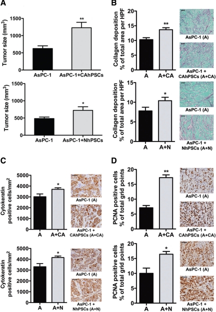
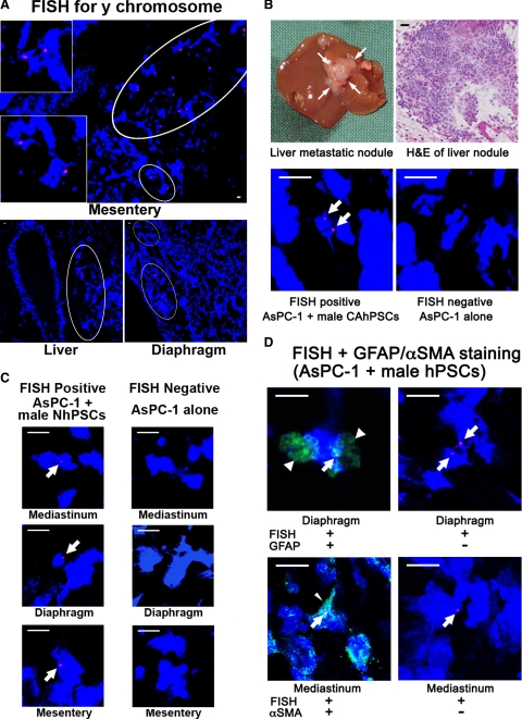
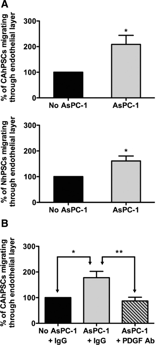
Pancreatic stellate cells (PSCs) produce the stromal reaction in pancreatic cancer (PC), and their interaction with cancer cells facilitates cancer progression. This study investigated the role of human PSCs (hPSCs) in the metastatic process and tumor angiogenesis using both in vivo (orthotopic model) and in vitro (cultured PSC and PC cells) approaches. A sex mismatch study (injection of male hPSCs plus female PC cells into the pancreas of female mice) was conducted to determine whether hPSCs accompany cancer cells to metastatic sites. Metastatic nodules were examined by fluorescent in situ hybridization for the presence of the Y chromosome. Angiogenesis was assessed by i) immunostaining tumors for CD31, an endothelial cell marker; and ii) quantifying human microvascular endothelial cell (HMEC-1) tube formation in vitro on exposure to conditioned media from hPSCs. Transendothelial migration was assessed in vitro by examining the movement of fluorescently labeled hPSCs through an endothelial cell monolayer. Human PSCs i) were found in multiple metastatic sites in each mouse injected with male hPSCs plus female PC cells; ii) increased CD31 expression in primary tumors from mice injected with MiaPaCa-2 and hPSCs and stimulated tube formation by HMEC-1 in vitro; and iii) exhibited transendothelial migration that was stimulated by cancer cells. Human PSCs accompany cancer cells to metastatic sites, stimulate angiogenesis, and are able to intravasate/extravasate to and from blood vessels.


Figures

Figure 1
Figure 1
Primary tumors in orthotopic model. A: Tumor size: Primary tumors in mice injected with AsPC-1 + CAhPSCs were significantly larger than tumors in mice injected with AsPC-1 alone (**P < 0.005; n = 14 per group). Similar results were observed in mice injected with AsPC-1 ± NhPSCs (*P < 0.05; n = 8 per group). B: Fibrosis in primary tumors: Representative Sirius Red–stained images (×400) showing significantly increased collagen deposition in primary tumors in the presence of CAhPSCs (**P < 0.001; n = 14 per group) and NhPSCs (*P < 0.05; n = 8 per group). C: Density of cancer cells: Representative images (×600) of sections immunostained for cytokeratin. The density of cytokeratin-positive cells was significantly higher in the primary tumors from mice injected with AsPC-1 + CAhPSCs or NhPSCs compared to tumors in mice injected with AsPC-1 alone (*P < 0.05; n = 14 per group and n = 8 per group, respectively). D: Cell proliferation in primary tumors: Immunostaining (×600) for PCNA, showing that the proportion of PCNA-positive cells was significantly increased in primary tumors from mice injected with AsPC-1 + CAhPSCs (**P < 0.0001; n = 14 per group) compared with that in tumors from mice injected with AsPC-1 alone. Similar results were found in mice injected with AsPC-1 ± NhPSCs (*P < 0.05; n = 8 per group). Scale bars = 50 μm.
Figure 2
Figure 2
Representative photomicrographs of metastatic nodules. H&E-stained sections of nodules (demarcated by arrows) in lung (×400), diaphragm (×200), liver (×200), and mesentery (×200). Scale bars = 100 μm.
Figure 3
Figure 3
Identification of male hPSCs in metastatic nodules. A: FISH for the Y chromosome: Representative photomicrographs (×200) showing Y chromosome–positive cells in metastatic nodules in the mesentery (insets are high-power views of the circled regions), liver, and diaphragm from mice injected with AsPC-1 + male hPSCs. B: Macroscopic appearance of a representative liver metastatic nodule from a mouse injected with AsPC-1 + male CAhPSCs and a photomicrograph (×400) of an H&E-stained section of a liver nodule. Lower panels show representative photomicrographs (×1000) of FISH for the Y chromosome (positive pink staining; arrows) in a liver metastatic nodule from a mouse injected with AsPC-1 + male CAhPSCs and no staining for the Y chromosome in a liver nodule from a mouse injected with AsPC-1 alone. C: FISH for the Y chromosome: Representative photomicrographs (×1000) showing positive pink staining (arrows) for the Y chromosome in metastatic nodules from mice injected with AsPC-1 + male NhPSCs (mediastinum, diaphragm, and mesentery). No such staining was observed in metastatic nodules of mice injected with AsPC-1 cells alone. D: Dual staining of cryosections: Representative photomicrographs of metastatic nodules stained for the Y chromosome using FISH (pink; arrows) and i) the PSC-selective marker GFAP (green; arrowheads) from mice injected with AsPC-1 + male NhPSCs or ii) the PSC activation marker αSMA (green; fine arrowhead) from mice injected with AsPC-1 + male CAhPSCs. Also shown are the corresponding negative controls for GFAP and αSMA where the FISH-positive sections were incubated with IgG1 or IgG2a respectively. Scale bars = 10 μm.
Figure 4
Figure 4
Angiogenesis assay. A: Effect of hPSCs on tube formation by HMEC-1 (×100): Conditioned media from CAhPSCs significantly stimulated tube formation by HMEC-1 cells (**P < 0.005; n = 4 different CAhPSC preparations; Ctrl = Control). Similar results were found with conditioned media from NhPSC preparations (**P < 0.01; n = 6 different NhPSC preparations). Scale bars = 50 μm. B: Effect of VEGF-neutralizing antibody on angiogenesis in vitro: Tube formation of HMEC-1 cells was significantly reduced in the presence of CAhPSC secretions pretreated with VEGF-neutralizing antibody (*P < 0.01, **P < 0.005; n = 3 different CAhPSC preparations).
Figure 5
Figure 5
Transendothelial migration of hPSCs. A: The proportion of CAhPSCs or NhPSCs migrating through an endothelial cell monolayer was significantly increased in the presence of AsPC-1 (*P < 0.05; n = 3 separate CAhPSC preparations and 5 separate NhPSC preparations). B: Effect of PDGF-neutralizing antibody on transendothelial migration: AsPC-1–stimulated transendothelial migration of CAhPSCs was prevented in the presence of PDGF-neutralizing antibody (*P < 0.05, **P < 0.01; n = 4 different CAhPSC preparations).

References

    1. Apte MV, Park S, Phillips PA, Santucci N, Goldstein D, Kumar RK, Ramm GA, Buchler M, Friess H, McCarroll JA, Keogh G, Merrett N, Pirola R, Wilson JS. Desmoplastic reaction in pancreatic cancer: role of pancreatic stellate cells. Pancreas. 2004;29:179–187. - PubMed
    1. Bachem MG, Schunemann M, Ramadani M, Siech M, Beger H, Buck A, Zhou S, Schmid-Kotsas A, Adler G. Pancreatic carcinoma cells induce fibrosis by stimulating proliferation and matrix synthesis of stellate cells. Gastroenterology. 2005;128:907–921. - PubMed
    1. Erkan M, Reiser-Erkan C, Michalski CW, Deucker S, Sauliunaite D, Streit S, Esposito I, Friess H, Kleeff J. Cancer-stellate cell interactions perpetuate the hypoxia-fibrosis cycle in pancreatic ductal adenocarcinoma. Neoplasia. 2009;11:497–508. - PMC - PubMed
    1. Hwang RF, Moore T, Arumugam T, Ramachandran V, Amos KD, Rivera A, Ji B, Evans DB, Logsdon CD. Cancer-associated stromal fibroblasts promote pancreatic tumor progression. Cancer Res. 2008;68:918–926. - PMC - PubMed
    1. Vonlaufen A, Joshi S, Qu C, Phillips PA, Xu Z, Parker NR, Toi CS, Pirola RC, Wilson JS, Goldstein D, Apte MV. Pancreatic stellate cells: partners in crime with pancreatic cancer cells. Cancer Res. 2008;68:2085–2093. - PubMed

Publication types