. 2010 Nov 15;70(22):9243-52.
doi: 10.1158/0008-5472.CAN-10-1250. Epub 2010 Oct 8.

MGMT-independent temozolomide resistance in pediatric glioblastoma cells associated with a PI3-kinase-mediated HOX/stem cell gene signature

Affiliations

MGMT-independent temozolomide resistance in pediatric glioblastoma cells associated with a PI3-kinase-mediated HOX/stem cell gene signature

Nathalie Gaspar et al. Cancer Res. .

Abstract

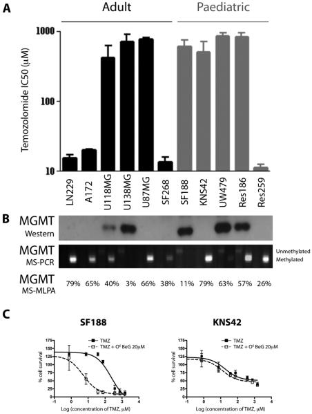
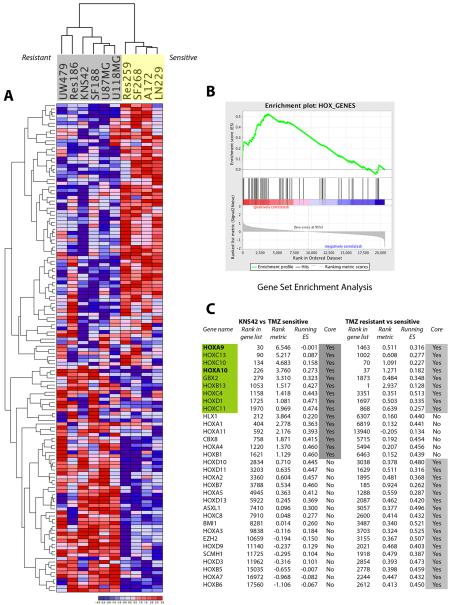
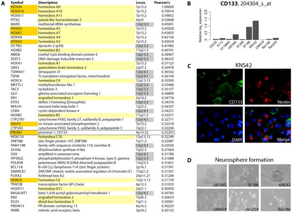
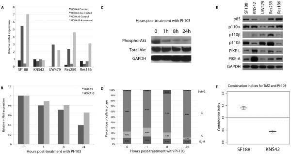
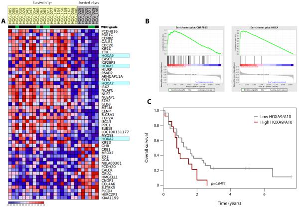
Sensitivity to temozolomide is restricted to a subset of glioblastoma patients, with the major determinant of resistance being a lack of promoter methylation of the gene encoding the repair protein DNA methyltransferase MGMT, although other mechanisms are thought to be active. There are, however, limited preclinical data in model systems derived from pediatric glioma patients. We screened a series of cell lines for temozolomide efficacy in vitro, and investigated the differential mechanisms of resistance involved. In the majority of cell lines, a lack of MGMT promoter methylation and subsequent protein overexpression were linked to temozolomide resistance. An exception was the pediatric glioblastoma line KNS42. Expression profiling data revealed a coordinated upregulation of HOX gene expression in resistant lines, especially KNS42, which was reversed by phosphoinositide 3-kinase pathway inhibition. High levels of HOXA9/HOXA10 gene expression were associated with a shorter survival in pediatric high-grade glioma patient samples. Combination treatment in vitro of pathway inhibition and temozolomide resulted in a highly synergistic interaction in KNS42 cells. The resistance gene signature further included contiguous genes within the 12q13-q14 amplicon, including the Akt enhancer PIKE, significantly overexpressed in the KNS42 line. These cells were also highly enriched for CD133 and other stem cell markers. We have thus shown an in vitro link between phosphoinositide 3-kinase-mediated HOXA9/HOXA10 expression, and a drug-resistant, progenitor cell phenotype in MGMT-independent pediatric glioblastoma.


Figures

Figure 1
Figure 1. Sensitivity of paediatric and adult glioma cell lines to TMZ and relationship to MGMT status
(A) Adult (LN229, A172, U118MG, U138MG, U87MG, SF268) and paediatric (SF188, KNS42, UW479, Res186, Res259) glioma cells were treated with TMZ, and cytotoxicity assessed by the SRB assay. IC50 values are plotted on a log10 scale. (B) Western blot for MGMT protein expression correlated with extent of promoter methylation as assessed by methylation-specific PCR and MLPA. In most cases expression correlates with TMZ resistance, with the exception of U87MG and KNS42 cells, which are hypermethylated, do not express the protein, and are resistant to TMZ. (C) SF188 and KNS42 cells were treated with MGMT substrate analogue O6-benzylguanine, demonstrating the MGMT-dependent nature of TMZ resistance in SF188, but not KNS42 cells. Growth inhibition was determined by the SRB assay. Concentration of TMZ is on a log10 scale.
Figure 2
Figure 2. Assessment of DNA mismatch repair and base excision repair pathways in paediatric and adult glioma cell lines
Western blot for proteins involved in (A) mismatch repair, and (B) base excision repair. Although deficiencies in MSH3 are noted in U138MG, UW479 and Res186, and there is some variability in PARP and XRCC1 expression, U87MG and KNS42 have normal expression of proteins involved in both pathways. TMZ-resistant cell lines are highlighted (*).
Figure 3
Figure 3. Expression profiling reveals a HOX / stem cell signature associated TMZ resistance in glioma cell lines
(A) Heatmap demonstrating hierarchical clustering of 135 differentially expressed genes between resistant (grey - UW479, Res186, KNS42, SF188, U87MG, U118MG) and sensitive (yellow – Res259, SF268, A172, LN229) high grade glioma cell lines. (B) Gene Set Enrichment Analysis highlighting co-ordinated differential expression of gene sets defined a priori. Enriched in TMZ resistant cell lines were the HOX_GENES set. (Enrichment Score = 0.54, nominal p = 0.01; FDR q = 0.403). (C) Ranked genes derived from GSEA of KNS42 versus TMZ sensitive cell lines, and all TMZ resistant versus sensitive lines. Genes are provided along with their rank in the total gene list, rank enrichment metric, and running enrichment score are provided for both analyses. Genes present within the core enrichment signature are highlighted in grey, with those genes present in both analyses highlighted in green. These genes which are also present in the HOX gene signature in Murat et al. (26), HOXA9 and HOXA10, are highlighted in bold.
Figure 4
Figure 4. Co-ordinated upregulation of HOX genes in primary glioblastomas and a striking degree of CD133 positivity on KNS42 cells
(A) Genes co-ordinately expressed with HOXA9 from the TCGA dataset (21) were determined by calculating Pearson’s correlation coefficients. All genes with values greater than 0.3 are listed, in rank order. Those highlighted in yellow are homeobox genes, genes in orange were also present in the HOX gene/self-renewal signature of Murat et al. (26), including PROM1 (CD133, box); genes highlighted in grey are found within the 12q13-q14 amplicon. (B) Affymetrix expression analysis of PROM1(CD133) mRNA levels in the panel of glioma cell lines, plotted as relative log2 expression. (C) Immunofluorescence assay demonstrating extensive expression of stem cell markers CD133 (green) and nestin (red) in KNS42 cells grown as a monolayer. (D) Neurosphere formation assay highlighting the tight three-dimensional spheroids formed by KNS42 cells in contrast to the other cell lines studied.
Figure 5
Figure 5. HOXA9/HOXA10 expression in KNS42 cells is driven by a lack of promoter methylation in a PI3-kinase-dependent manner
(A) Relative mRNA expression levels of HOXA9 and HOXA10 before and after treatment with 5-Aza-2′-deoxycytidine in paediatric glioma cell lines. An absence of expression changes after 5-Aza-2′-deoxycytidine treatment in KNS42 cells is indicative of an absence of constitutive promoter methylation. (B) HOXA9/HOXA10 expression is reduced by treatment with PI-103 in KNS42 cells in a time-dependent manner. Treatment with the dual PI3-kinase/mTOR inhibitor PI-103 at 5xIC50 for 1, 8 and 24 hours led to a reduction in expression of both HOXA9 and HOXA10 in KNS42 cells as early as 1 hour post-treatment. (C) Western blot analysis of phospho- and total Akt after treatment of KNS42 cells with PI-103 at 5xIC50 for 1, 8 and 24 hours. Inhibition of PI3K signalling is observed at the earliest timepoint (D) Cell cycle analysis of KNS42 cells after treatment with PI-103 at 5xIC50 for 1, 8 and 24 hours. There was no G1 arrest evident at the early timepoints at which reduced HOXA9/HOXA10 expression was observed. (E) Western blot analysis of PI3-kinase regulatory and catalytic subunits and enhancers in paediatric glioma cells. Expression of the specific Akt enhancer proteins PIKE-A and PIKE-L were considerably elevated in KNS42 cells compared with other paediatric glioma cells. (F) Synergistic interaction of PI-103 and TMZ. Co-treatment with PI-103 and TMZ resulted in a high degree of synergy in MGMT-independent KNS42 cells (combination index = 0.43) as calculated by the median effect analysis. By contrast, SF188 cells showed an antagonistic interaction (combination index = 1.401).
Figure 6
Figure 6. High levels of HOXA gene expression are associated with shorter survival in paediatric high grade glioma patients
(A) Heatmap representing differentially expressed genes between short (<1 year, yellow) and long-term (>3 years, grey) survivors. WHO grade IV (black) and III (green) tumours are indicated. HOXA genes are highlighted in light blue. (B) GSEA analysis demonstrating significant enrichment of genes at the chromosome 7p15 locus (Enrichment Score = 0.68, nominal p < 0.001, FDR q = 0.930); and of HOXA genes in particular (Enrichment Score = 0.90, nominal p < 0.001, FDR q < 0.001), upregulated in short term survivors. (C) Kaplan-Meier plot demonstrating a significantly shorter survival of patients with high levels of HOXA9/HOXA10 gene expression (p=0.0453, log-rank test).

References

    1. Mrugala MM, Chamberlain MC. Mechanisms of disease: temozolomide and glioblastoma--look to the future. Nat Clin Pract Oncol. 2008;5:476–86. - PubMed
    1. Stupp R, Mason WP, van den Bent MJ, Weller M, Fisher B, Taphoorn MJ, et al. Radiotherapy plus concomitant and adjuvant temozolomide for glioblastoma. N Engl J Med. 2005;352:987–96. - PubMed
    1. Newlands ES, Blackledge GR, Slack JA, Rustin GJ, Smith DB, Stuart NS, et al. Phase I trial of temozolomide (CCRG 81045: M&B 39831: NSC 362856) Br J Cancer. 1992;65:287–91. - PMC - PubMed
    1. Hegi ME, Diserens AC, Gorlia T, Hamou MF, de Tribolet N, Weller M, et al. MGMT gene silencing and benefit from temozolomide in glioblastoma. N Engl J Med. 2005;352:997–1003. - PubMed
    1. Esteller M, Garcia-Foncillas J, Andion E, Goodman SN, Hidalgo OF, Vanaclocha V, et al. Inactivation of the DNA-repair gene MGMT and the clinical response of gliomas to alkylating agents. N Engl J Med. 2000;343:1350–4. - PubMed

Publication types

MeSH terms

Associated data