. 2010 Nov 3;102(21):1637-52.
doi: 10.1093/jnci/djq361. Epub 2010 Oct 8.

Prolonged drug selection of breast cancer cells and enrichment of cancer stem cell characteristics

Affiliations

Prolonged drug selection of breast cancer cells and enrichment of cancer stem cell characteristics

Anna Maria Calcagno et al. J Natl Cancer Inst. .

Abstract

Background: Cancer stem cells are presumed to have virtually unlimited proliferative and self-renewal abilities and to be highly resistant to chemotherapy, a feature that is associated with overexpression of ATP-binding cassette transporters. We investigated whether prolonged continuous selection of cells for drug resistance enriches cultures for cancer stem-like cells.

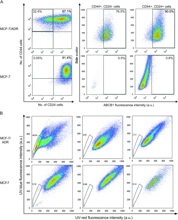
Methods: Cancer stem cells were defined as CD44+/CD24⁻ cells that could self-renew (ie, generate cells with the tumorigenic CD44+/CD24⁻ phenotype), differentiate, invade, and form tumors in vivo. We used doxorubicin-selected MCF-7/ADR cells, weakly tumorigenic parental MCF-7 cells, and MCF-7/MDR, an MCF-7 subline with forced expression of ABCB1 protein. Cells were examined for cell surface markers and side-population fractions by microarray and flow cytometry, with in vitro invasion assays, and for ability to form mammospheres. Xenograft tumors were generated in mice to examine tumorigenicity (n = 52). The mRNA expression of multidrug resistance genes was examined in putative cancer stem cells and pathway analysis of statistically significantly differentially expressed genes was performed. All statistical tests were two-sided.

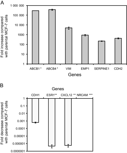
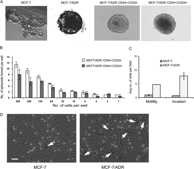
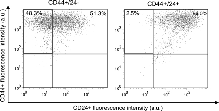
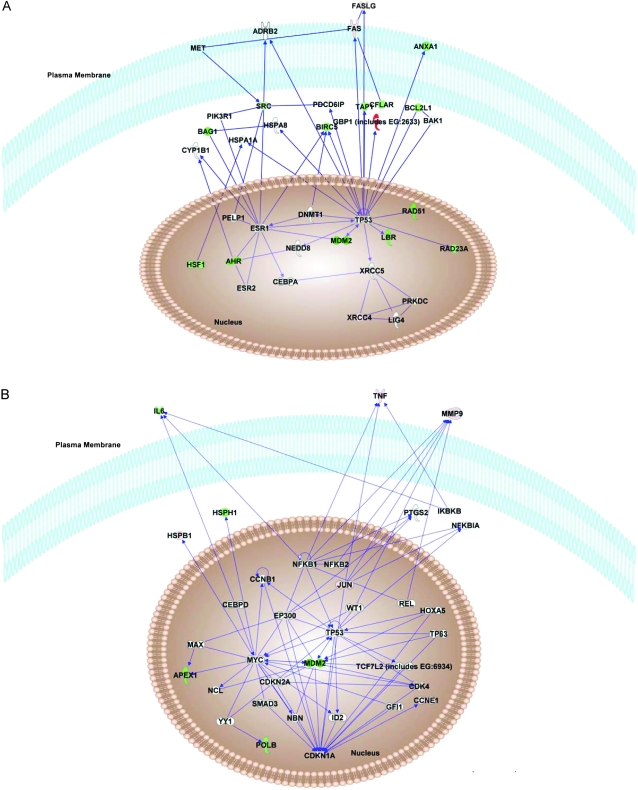
Results: Pathway analysis showed that MCF-7/ADR cells express mRNAs from ABCB1 and other genes also found in breast cancer stem cells (eg, CD44, TGFB1, and SNAI1). MCF-7/ADR cells were highly invasive, formed mammospheres, and were tumorigenic in mice. In contrast to parental MCF-7 cells, more than 30% of MCF-7/ADR cells had a CD44+/CD24⁻ phenotype, could self-renew, and differentiate (ie, produce CD44+/CD24⁻ and CD44+/CD24+ cells) and overexpressed various multidrug resistance-linked genes (including ABCB1, CCNE1, and MMP9). MCF-7/ADR cells were statistically significantly more invasive in Matrigel than parental MCF-7 cells (MCF-7 cells = 0.82 cell per field and MCF-7/ADR = 7.51 cells per field, difference = 6.69 cells per field, 95% confidence interval = 4.82 to 8.55 cells per field, P < .001). No enrichment in the CD44+/CD24⁻ or CD133+ population was detected in MCF-7/MDR.

Conclusion: The cell population with cancer stem cell characteristics increased after prolonged continuous selection for doxorubicin resistance.


Figures

Figure 1
Figure 1
Differential gene expression in long-term doxorubicin-selected MCF-7/ADR cells and parental MCF-7 cells. A) Six genes with higher expression in MCF-7/ADR cells than in parental MCF-7 cells. B) Four genes with lower expression in MCF-7/ADR cells than in parental MCF-7 cells. Real-time quantitative reverse transcription–polymerase chain reaction was used to measure the mRNA expression levels of genes with statistically significantly different expression, as detected in the microarray analysis, between MCF-7/ADR and MCF-7 cells. * = Not detected in parental MCF-7 cells; ** = not detected in MCF-7/ADR cells; *** = not detected in either cell line. Data are the average fold change values in MCF-7/ADR cells compared with MCF-7 cells and are from at least two independent experiments, with at least two replicates per gene. Error bars = 95% confidence intervals.
Figure 2
Figure 2
Analysis of mammary stem cell markers in cultures of MCF-7/ADR cells and parental MCF-7 cells. We used CD44, CD24, and ABCB1 expression as stem cell markers and transport of Hoechst 33342 dye to indicate ABCB1 function. Cells were sorted for these markers by flow cytometry. A) Analysis of the cell surface expression of CD44 and CD24 protein on MCF-7 parental cells and multistep doxorubicin-selected MCF-7/ADR cells. CD44 and CD24 protein were detected with fluorochrome-conjugated antibodies. Flow cytometry was used to sort cells into CD44+/CD24− and CD44+/CD24+ populations, and the expression of ABCB1 protein was assessed in each population, also by flow cytometry. Histograms from a representative experiment of a total of four experiments are shown. Percentages indicate the amount of cells in that quadrant. Side scatter is reflected and refracted light intensity related to cell granularity and complexity. B) Analysis of the side-population cell fraction in MCF-7 and MCF-7/ADR cell cultures. Left) Hoechst 33342 dye treatment only. Middle) Verapamil treatment. Right) Cyclosporine A treatment. All cells were incubated with Hoechst 33342 dye, which was excited at 357 nm and its fluorescence was analyzed at two wavelengths (blue = 402–446 nm; and red = 650–670 nm), both in linear mode. Cells were incubated with 50 μM verapamil or 10 μM cyclosporine A before addition of Hoechst dye; dead cells were excluded by incubation with 7-AAD (20 μg/mL) immediately before analysis. Cell aggregates were discarded from the analysis by doublet discrimination. Side-population cells are shown by the outlined area. a.u. = arbitrary units.
Figure 3
Figure 3
Characterization of MCF-7 and MCF-7/ADR cells in three-dimensional culture. Mammosphere formation by unsorted and sorted (CD44+/CD24− and CD44+/CD24+) cell populations of MCF-7/ADR cells and parental MCF-7 cells was assessed by the limiting dilution assay. A) Morphologic appearance of mammospheres. A representative mammosphere from a total of 50 countable mammospheres from each experimental condition is shown. B) Spheroid formation by sorted cells in anchorage-independent culture. Long-term spheroid assays were performed by limiting dilution with 500 cells to one cell per well of a 96-well low-binding plate. The number of colonies was scored at the end of 14 days. This experiment was performed twice, with six wells per cell dilution. Error bars = 95% confidence intervals. C) Motility and invasiveness of MCF-7/ADR cells and parental MCF-7 cells. Cell culture inserts were used for both assays. Membranes coated with Matrigel were used for invasion assays, and membranes without a Matrigel coating were used for motility assays. For each insert, cells in five different ×10 fields were counted. Cell numbers from two experiments were averaged. Number of replicates (wells) for each bar is four. Differences in motility or invasiveness were compared with a two-sided t test (both P < .001). D) Representative inserts from invasion assays for MCF-7/ADR cells and parental MCF-7 cells. White circles are pores. MCF-7 cell in the field captured on the left contains only one cell (arrow). MCF-7/ADR cells in the right panel are more numerous and elongated, as indicated by arrows, with a mesenchymal appearance. Cultures were photographed at a magnification of ×40. Scale bar = 50 μm.
Figure 4
Figure 4
Self-renewal and differentiation abilities of sorted MCF-7/ADR cell population. Self-renewal was defined as maintenance of cells with cancer stem cell markers, CD44+/CD24–. Differentiation was defined as loss of these markers and appearance of CD44+/CD24+ double-positive populations. Cultures of MCF-7/ADR cells were sorted by flow cytometry into CD44+/CD24− or CD44+/CD24+ populations, each cell type was grown as mammospheres for 16 days, and then cells in the mammospheres were examined for cell surface expression of CD44 and CD24 by flow cytometry. Percentages in a quadrant indicate the relative numbers of cells in that quadrant at the end of the experiment. In both panels, upper left quadrant = CD44+/CD24– cells; upper right = CD44+/CD24+ cells; lower left = CD44–/CD24− cells, and lower right = CD44−/CD24+ cells. Data are from one experiment with duplicate samples; a total of two experiments were performed.
Figure 5
Figure 5
Analysis of gene expression in CD44+/CD24− MCF-7/ADR cell population by TaqMan Low-Density Array and Ingenuity Pathway analysis. Genes in CD44+/CD24− MCF-7/ADR cells with expression levels that were greater than twofold higher or lower than their expression in CD44+/CD24+ MCF-7/ADR cells (Supplementary Table 7, available online) were analyzed by Ingenuity Pathways software to identify any biological relationships among these genes. A) Highest ranked pathway. B) Second highest ranked pathway. Green shapes = decreased gene expression; red shapes = increased gene expression. Pathways were drawn by use of the Path Designer feature in Ingenuity Pathways Analysis software, version 8.0 (Ingenuity).

Similar articles

Cited by

References

    1. Gottesman M, Fojo T, Bates S. Multidrug resistance in cancer: role of ATP-dependent transporters. Nat Rev Cancer. 2002;2(1):48–58. - PubMed
    1. Calcagno AM, Kim IW, Wu CP, Shukla S, Ambudkar SV. ABC drug transporters as molecular targets for the prevention of multidrug resistance and drug-drug interactions. Curr Drug Deliv. 2007;4(4):324–333. - PubMed
    1. Bertolini G, Roz L, Perego P, et al. Highly tumorigenic lung cancer CD133+ cells display stem-like features and are spared by cisplatin treatment. Proc Natl Acad Sci U S A. 2009;106(38):16281–16286. - PMC - PubMed
    1. Creighton CJ, Li X, Landis M, et al. Residual breast cancers after conventional therapy display mesenchymal as well as tumor-initiating features. Proc Natl Acad Sci U S A. 2009;106(33):13820–13825. - PMC - PubMed
    1. Fillmore CM, Kuperwasser C. Human breast cancer cell lines contain stem-like cells that self-renew, give rise to phenotypically diverse progeny and survive chemotherapy. Breast Cancer Res. 2008;10(2):R25. - PMC - PubMed

Publication types

MeSH terms