. 2010 Oct 1;9(19):3921-32.
doi: 10.4161/cc.9.19.13139. Epub 2010 Oct 25.

Nitrogen anabolism underlies the importance of glutaminolysis in proliferating cells

Affiliations

Nitrogen anabolism underlies the importance of glutaminolysis in proliferating cells

Meng Meng et al. Cell Cycle. .

Abstract

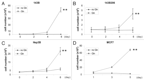
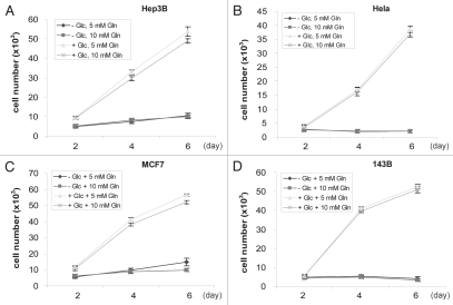
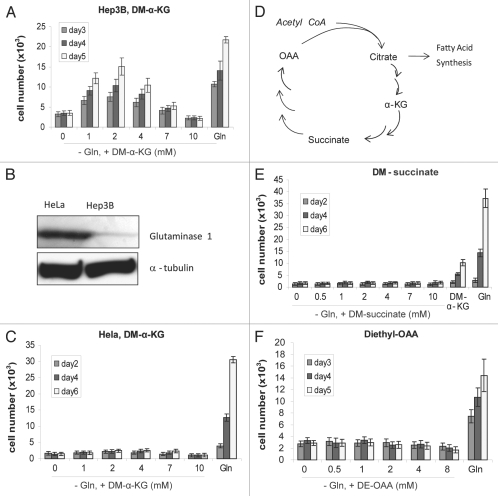
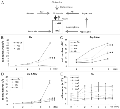
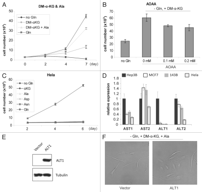
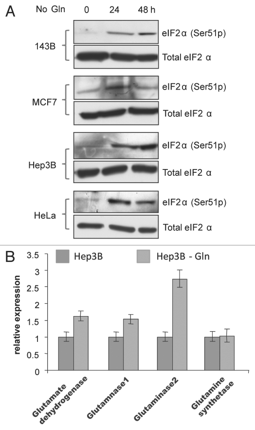
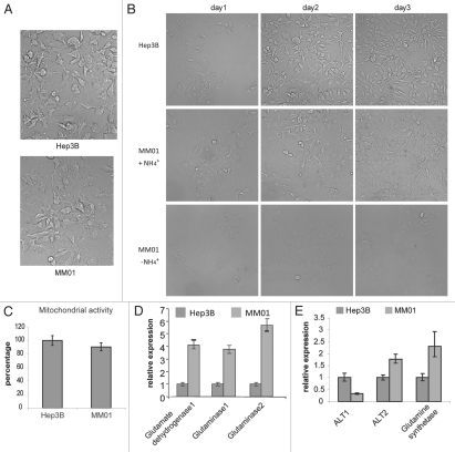
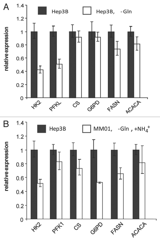
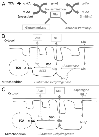
Glutaminolysis and Warburg effect are the two most noticeable metabolic features of tumor cells whereas their biological significance in cell proliferation remains elusive. A widely accepted current hypothesis is that tumor cells use glutamine as a preferred carbon source for energy and reducing power, which has been used to explain both glutaminolysis and the Warburg effect. Here we provide evidence to show that supplying nitrogen, not the carbon skeleton, underlies the major biological importance of glutaminolysis for proliferating cells. We show alternative nitrogen supplying mechanisms rescue cell proliferation in glutamine-free media. Particularly, we show that ammonia is sufficient to maintain a long-term survival and proliferation of Hep3B in glutamine-free media. We also observed that nitrogen source restriction repressed carbon metabolic pathways including glucose utilization. Based on these new observations and metabolic pathways well established in published literature, we propose an alternative model that cellular demand for glutamate as a key molecule in nitrogen anabolism is the driving force of glutaminolysis in proliferating cells. Our model suggests that the Warburg effect may be a metabolic consequence secondary to the nitrogen anabolism.


Figures

Figure 1
Figure 1
All cell lines tested require Gln for proliferation. Cells were seeded in Gln-free DMEM supplemented with 0 or 4 mM Gln. Cell numbers were analyzed every 2 days and mean values were shown. Representative growth curves of 143B (A), 143B206 (B), Hep3B (C) and MCF7 (D) cells are shown. Error bars indicate SE of ≥4 replicates.
Figure 2
Figure 2
All cell lines tested require glucose for proliferation. Cells were seeded in Gln-free DMEM supplemented with 0 or 4.5 g/L Glc and 5 or 10 mM Gln, respectively. Cell numbers were analyzed every 2 days and mean values were shown. Representative growth curves of Hep3B (A), Hela (B), MCF7 (C) and 143B (D) cells are shown. Error bars indicate SE of ≥4 replicates.
Figure 3
Figure 3
Rescuing effect of DM-α-KG is not based on anaplerosis. Increasing concentrations of DM-α-KG were used in cell culture. Mean value of cell number was shown. Error bars indicate SE of ≥4 replicates. (A) Biphasic effect of DM-α-KG on proliferation of Hep3B cells. (B) Protein levels of glutaminase in HeLa and Hep3B cells. 80 µg of total proteins were loaded onto a SDS page. Glutaminase levels were determined by western blotting. α-tubulin was used as a loading control. (C) α-KG failed to rescue HeLa cell proliferation in Glu-free media. (D) Simplified drawing showing the major anaplerotic metabolites of TCA cycle. In addition to α-KG, succinate and OAA have anaplerotic roles. (E and F) Growth curve of Hep3B cells in Gln-free DMEM supplemented with either 1–8 mM of diethyl-OAA or dimethyl-succinate. DMEM and complete DMEM (+Gln) were respectively set as controls.
Figure 4
Figure 4
Alternative nitrogen sources rescue Hep3B. (A) Nitrogen metabolic pathways relevant to this study. Glutaminolysis and the metabolic pathways to utilize Ala, Asp, Asn and ammonia as nitrogen sources were shown. (B–D) Growth curve of Hep3B cells in Gln-free DMEM supplemented with 4 mM Asns or Asp (B), with 4 mM Ala (C), with 0.8 mM ammonia or 4 mM Glu (D). For (B–D) cells seeded in Gln-free DMEM were used as control. Cell number was analyzed every 2 or 3 days and mean values were shown. (E) Dose curve of Hep3B cells in various concentrations of Glu.
Figure 5
Figure 5
Rescuing effects of α-KG, Ala and Asp depend on transamination. (A) Synergetic rescuing effect of Ala and α-KG. Growth curve of Hep3B cells in Gln-free DMEM supplemented with DM-α-KG (2 mM) or a combination of DM-α-KG (2 mM) and Ala (4 mM). Mean value of cell number was shown with plotted lines. Error bars indicate SE of ≥4 replicates. (B) Presence of transaminase inhibitor impairs the rescuing effects of α-KG. AOAA was used at 0.1 and 0.2 mM. Data represent percentage of cell numbers compared with those in normal media at day 4. Cell numbers with normal media were set as 100%. (C) HeLa cells cannot be rescued by either α-KG or alternative nitrogen sources. (D) q-RT-PCR analysis showing ALT1 and AST1 levels are associated with the rescuing effect. (E) Exogenous expression of ALT1 in HeLa cells. (F) Expression of ALT1 in combination with Ala and α-KG rescues HeLa proliferation. HeLa cells transfected with vector or ALT1-expressing plasmids were grown in Gln-free media for 7 days. Representative images were taken (200×).
Figure 6
Figure 6
Gln depletion triggers amino acid deprivation response and adaptive expression of genes important for Glu metabolism. (A) Immunoblotting analysis of phosphorylated-(Ser51) eIF2α, a biomarker of amino acid deprivation stress response. Total eIF2α was detected as control. (B) Gln depletion upregulates genes involved in nitrogen metabolism. Cells were cultured in media with or without Gln for 48 h, and total RNA samples were prepared. The mRNA levels of genes encoding major nitrogen metabolic enzymes were analyzed by real-time RT-PCR.
Figure 7
Figure 7
Establishment of cell lines growing in ammonia-containing media. (A) Morphology of MM01 and Hep3B (200×). (B) Ammonia is sufficient to support long-term proliferation of Hep3B in Gln-free media. Hep3B cells were seeded in complete media (three top parts) as control. MM01 cells were seeded in Gln-free media with ammonia (three middle parts). Withdrawal of ammonia suppressed proliferation (three bottom parts). All micrographs were taken at 100× magnification. (C) Mitochondrial activity of Hep3B and MM01 as determined by MTT assays. (D) Adaptive expression of genes encoding major enzymes involved in Glu homeostasis. (E) Change of expression of ALT1/2 and Gln synthetase were determined by real-time RT-PCR. Data indicate that MM01 growing on ammonia-expressing genes facilitate nitrogen flux to the synthesis of Ala (ALT2) and Gln (glutamine synthetase).
Figure 8
Figure 8
Nitrogen restriction represses the utilization of glucose. Relative mRNA levels of genes involved in glucose utilization were determined by qRT-PCR. Hep3B cells grown in normal DMEM medium were set as reference. (A) Relative gene expression levels in Hep3B cells treated by Gln-free DMEM for 12 hours. (B) Relative gene expression level in MM01 cells grown in Gln-free DMEM supplemented with 0.4 mM (NH4)2SO4. HK2, hexokinase 2; PFK1, phosphofructokinase 1; CS, citrate synthase; G6PD, glucose-6-phosphate dehydrogenase; FASN, fatty acid synthase; ACACA, acetyl-CoA carboxylase.
Figure 9
Figure 9
Proposed role of glutaminolysis in nitrogen anabolism in proliferating cells. (A) Central roles of interconversion between α-KG and Glu in nitrogen metabolism. The interconversion between α-KG and Glu in cytoplasmic provides a mechanism to collect amino groups from excessive amino acids and redistribute them to produce amino acids on demand. An adequate pool of cytoplasmic α-KG and Glu is essential for this function. In proliferating cells, Glu is actively consumed for biosynthesis. Because the uptake of Glu is limited by the capacity of cell surface transporters, the loss of cytoplamic gluatamate is mainly replenished by glutaminolysis. Excessive amino acids may replenish the Glu pool, contingent on the transaminase acitivity. Excessive α-KG inhibits transamination from Glu to other α-KAs, impairing the biosynthesis of amino acids in demand. (B) Glutaminolysis and Gln-Glu/Asp exchange model. When Gln is available, Gln enters mitochondria and is hydrolyzed to Glu. In addition to being consumed by the Krebs cycle, mitochondrial Glu can exported outside to maintain the homeostasis of the cytoplasmic Glu-α-KG pool or used to generate Asp for export. (C) Recycling of ammonia for nitrogen anabolism by specific cell types. When Gln is scarce, some types of cells may use ammonia (in media or from catabolism of amino acids such as Asn) to synthesize Glu. This pathway depends on the GLUD activity. α-KA, α-keto acids; α-AA, α-amino acids.

References

    1. Warburg O. On the origin of cancer cells. Science. 1956;123:309–314. - PubMed
    1. Klimberg VS, McClellan JL, Organ CH., Jr Honorary Lectureship. Glutamine, cancer and its therapy. Am J Surg. 1996;172:418–424. - PubMed
    1. Pavlides S, Whitaker-Menezes D, Castello-Cros R, Flomenberg N, Witkiewicz AK, Frank PG, et al. The reverse Warburg effect: Aerobic glycolysis in cancer associated fibroblasts and the tumor stroma. Cell Cycle. 2009;8:3984–4001. - PubMed
    1. Medina MA. Glutamine and cancer. J Nutr. 2001;131:2539–2542. - PubMed
    1. Souba WW. Glutamine and cancer. Ann Surg. 1993;218:715–728. - PMC - PubMed

Publication types