Review
. 2011 Jan;7(1):2-11.
doi: 10.4161/auto.7.1.13044. Epub 2011 Jan 1.

The emerging role of autophagy in the pathophysiology of diabetes mellitus

Affiliations
Review

The emerging role of autophagy in the pathophysiology of diabetes mellitus

Claudio D Gonzalez et al. Autophagy. 2011 Jan.

Abstract

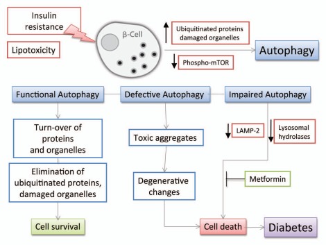
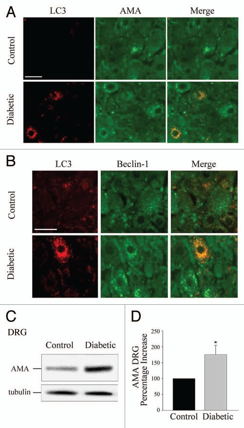
An emerging body of evidence supports a role for autophagy in the pathophysiology of type 1 and type 2 diabetes mellitus. Persistent high concentrations of glucose lead to imbalances in the antioxidant capacity within the cell resulting in oxidative stress-mediated injury in both disorders. An anticipated consequence of impaired autophagy is the accumulation of dysfunctional organelles such as mitochondria within the cell. Mitochondria are the primary site of the production of reactive oxygen species (ROS), and an imbalance in ROS production relative to the cytoprotective action of autophagy may lead to the accumulation of ROS. Impaired mitochondrial function associated with increased ROS levels have been proposed as mechanisms contributing to insulin resistance. In this article we review and interpret the literature that implicates a role for autophagy in the pathophysiology of type 1 and type 2 diabetes mellitus as it applies to β-cell dysfunction, and more broadly to organ systems involved in complications of diabetes including the cardiovascular, renal and nervous systems.


Figures

Figure 1
Figure 1
Autophagosome formation in pancreatic β-cells. Electron microscopy analysis reveals enhanced autophagosome formation in β-cells of 20-week-old mice fed a high-fat diet for 12 weeks (from 8 to 20 weeks of age). (A) Graph: enhanced formation of autophagosomes in β-cells of high-fat diet-fed C57BL/6 mice (HF) compared with standard diet-fed group (STD). Density of autophagosomes was calculated, and data are presented as mean ± SEM of three mice. *p < 0.01 versus controls. (B) FFA but not high glucose can induce autophagy in INS-1 cells. For control, cells were incubated in RPMI 1640 supplemented with FCS and amino acids. For starvation, cells were incubated for 2 h in Hank's buffer free of serum and amino acids. So that accumulation of LC3-II could be visualized, the lysosome inhibitors pepstatin A (10 µg/ml) and E-64-d (10 µg/ml) were added to the culture medium for the last 2 h period unless otherwise indicated. Adapted from reference , with permission.
Figure 2
Figure 2
Accumulation of ubiquitin aggregates in pancreatic β-cells of Atg7Δ β-cell mice. Pancreatic sections were subjected to immunohistochemistry using anti-ubiquitin antibodies. Several islet cells show accumulation of large protein aggregates containing ubiquitin (dark-brown spots). Adapted from reference , with permission.
Figure 3
Figure 3
Electron microscopy of a normal human β-cell (A), an apoptotic type 2 diabetic β-cell with marked chromatin condensation (B), and a dead type 2 diabetic β-cell with evidence of massive vacuoles overload (C). N, nucleus; insulin granules. Magnification x10,000 (A and C), x7,000 (B). Adapted from reference , with permission.
Figure 4
Figure 4
Schematic diagram of the effects of insulin resistance on β-cell autophagy. Various stresses caused by peripheral insulin resistance can induce polyubiquitination of proteins and accumulation of damaged organelles as cytoplasmic aggregates in pancreatic β-cells. Ubiquitinated proteins and damaged organelles, which otherwise form toxic aggregates, are effectively cleared by autophagy. Thus, autophagy can be viewed as a defense mechanism against cellular damage in β-cells. Elevated free-fatty acids also stimulate β-cell autophagy by gradual decrease in the level of phospho-mTOR under the state of insulin resistance. Interestingly, lipotoxicity induces impaired autophagy in human β-cells with reduction of LAMP-2 and lysosomal hydrolases leading to vacuole accumulation and cell death.
Figure 5
Figure 5
Diabetic rats demonstrate increased levels of anti-LC3 and anti-mitochondria (AMA) immunoreactivity that colocalize in dorsal root ganglion (DRG) neurons. (A) IHC detection of LC3 and colocalization with the mitochondrial AMA signal in DRG neurons from control and diabetic rats. (B) IHC detection of Beclin 1, LC3 and their colocalization in DRG neurons of normal, control and diabetic rats. (C) Representative immunoblot of mitochondrial (AMA) in DRG homogenates from normal (control) and diabetic rats. (D) Histogram of densitometric scannings of immunoblots of mitochondria (AMA) in DRG homogenates from control and diabetic rat DRG neurons. The normal control was set to 100%. Scale bar, 50 µm. *p < 0.05. Adapted from reference , with permission.

References

    1. Levine B, Kroemer G. Autophagy in the pathogenesis of disease. Cell. 2008;132:27–42. - PMC - PubMed
    1. Kim EH, Sohn S, Kwon HJ, Kim SU, Kim MJ, Lee SJ, Choi KS. Sodium selenite induces superoxide-mediated mitochondrial damage and subsequent autophagic cell death in malignant glioma cells. Cancer Res. 2007;67:6314–6324. - PubMed
    1. Park KJ, Lee SH, Kim TI, Lee HW, Lee CH, Kim EH, et al. 3: A human scfv antibody against trail receptor 2 induces autophagic cell death in both trail-sensitive and trail-resistant cancer cells. Cancer Res. 2008;67:7317–7334. - PubMed
    1. Pyo JO, Jang MH, Kwon YK, Lee HJ, Jun JI, Woo HN, et al. Essential roles of atg5 and FADD in autophagic cell death: Dissection of autophagic cell death into vacuole formation and cell death. J Biol Chem. 2007;280:20722–20729. - PubMed
    1. Boya P, Gonzalez-Polo RA, Casares N, Perfettini JL, Dessen P, Larochette N, et al. Inhibition of macroautophagy triggers apoptosis. Mol Cell Biol. 2005;25:1025–1040. - PMC - PubMed

Publication types

MeSH terms