

TLRs, NF-κB, JNK, and Liver Regeneration
- PMID: 20936148
- PMCID: PMC2948885
- DOI: 10.1155/2010/598109
TLRs, NF-κB, JNK, and Liver Regeneration
Abstract
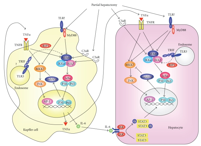
While hepatocytes rarely undergo proliferation in normal livers, they quickly induce proliferation in response to loss of liver mass by toxin or inflammation-induced hepatocyte injury, trauma, or surgical resection, leading to a restoration of liver mass to its original size. Recent studies suggest that Toll-like receptor (TLR) signaling participates in this regenerative response. Myeloid differentiation factor (MyD88), a common adaptor molecule in the TLR, IL-1 and IL-18 receptor signaling, plays a key role, at least, in the early phase of liver regeneration. Currently, definite ligands which bind to TLRs and initiate this process are still unclear. TLRs stimulated by their corresponding ligands, as well as tumor necrosis factor (TNF) receptors (TNFRs), can activate downstream signal molecules, including transcription factor nuclear factor (NF)-κB and c-Jun N-terminal kinase (JNK). Previous studies have revealed the important role of TNF receptor signaling, NF-κB, and JNK in liver regeneration by using hepatocyte-specific gene-modified animals. This review will summarize the current knowledge of TLR signaling and their related molecules in liver regeneration. We will also discuss whether modulating these factors may become new therapeutic strategies to promote liver regeneration in various clinical situations.
Figures
References
-
- Michalopoulos GK, DeFrances MC. Liver regeneration. Science. 1997;276(5309):60–65. - PubMed
-
- Taub R. Liver regeneration: from myth to mechanism. Nature Reviews Molecular Cell Biology. 2004;5(10):836–847. - PubMed
-
- Fausto N, Campbell JS, Riehle KJ. Liver regeneration. Hepatology. 2006;43(2, supplement 1):S45–S53. - PubMed
LinkOut - more resources
Full Text Sources
Research Materials
Miscellaneous
