. 2011 Feb;121(2):193-205.
doi: 10.1007/s00401-010-0756-0. Epub 2010 Oct 10.

Patterns of microRNA expression in normal and early Alzheimer's disease human temporal cortex: white matter versus gray matter

Affiliations

Patterns of microRNA expression in normal and early Alzheimer's disease human temporal cortex: white matter versus gray matter

Wang-Xia Wang et al. Acta Neuropathol. 2011 Feb.

Abstract

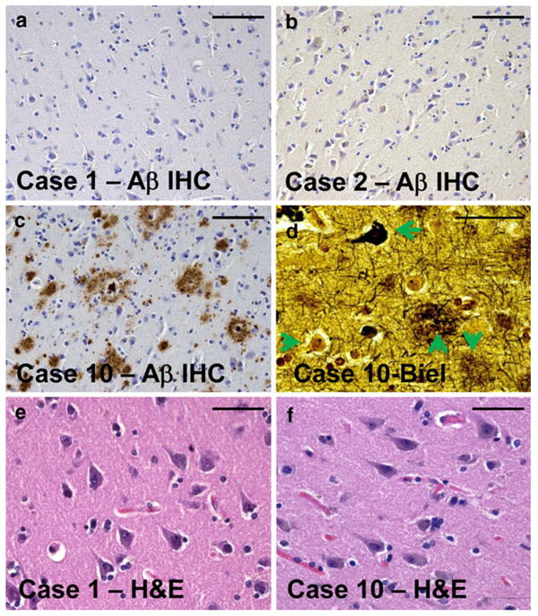
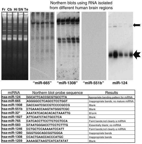
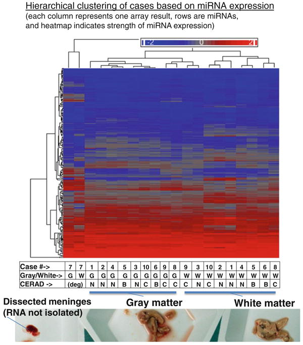
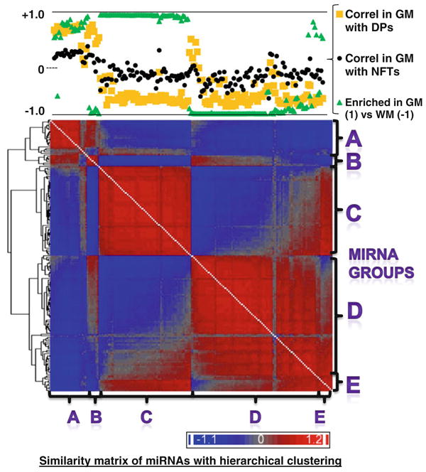
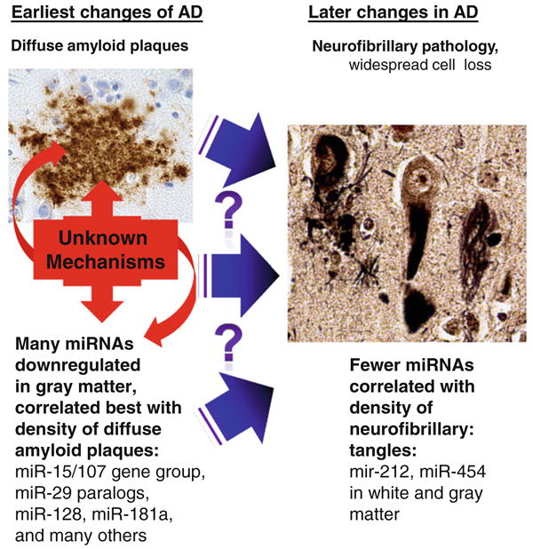
MicroRNA (miRNA) expression was assessed in human cerebral cortical gray matter (GM) and white matter (WM) in order to provide the first insights into the difference between GM and WM miRNA repertoires across a range of Alzheimer's disease (AD) pathology. RNA was isolated separately from GM and WM portions of superior and middle temporal cerebral cortex (N = 10 elderly females, postmortem interval < 4 h). miRNA profiling experiments were performed using state-of-the-art Exiqon(©) LNA-microarrays. A subset of miRNAs that appeared to be strongly expressed according to the microarrays did not appear to be conventional miRNAs according to Northern blot analyses. Some well-characterized miRNAs were substantially enriched in WM as expected. However, most of the miRNA expression variability that correlated with the presence of early AD-related pathology was seen in GM. We confirm that downregulation of a set of miRNAs in GM (including several miR-15/107 genes and miR-29 paralogs) correlated strongly with the density of diffuse amyloid plaques detected in adjacent tissue. A few miRNAs were differentially expressed in WM, including miR-212 that is downregulated in AD and miR-424 which is upregulated in AD. The expression of certain miRNAs correlates with other miRNAs across different cases, and particular subsets of miRNAs are coordinately expressed in relation to AD-related pathology. These data support the hypothesis that patterns of miRNA expression in cortical GM may contribute to AD pathogenetically, because the aggregate change in miRNA expression observed early in the disease would be predicted to cause profound changes in gene expression.


Figures

Fig. 1
Fig. 1
Representative photomicrographs from the superior and middle temporal cortical gyri of individuals sampled for the current study. Tissue sections for histopathology (Table 1) were immediately adjacent to the tissue samples used for RNA isolation. Amyloid plaques are stained using Aβ immunohistochemistry (IHC) as shown in ac. Note that in cases 1 and 2 (a, b), there is no observable Aβ plaques. By contrast, case 10 (c, d, f) had the highest densities of Alzheimer's disease (AD)-related pathology. Bielschowsky (Biel) silver stain (d) confirms the presence in case 10 of diffuse amyloid plaques (downward arrow), neuritic amyloid plaques (upward arrow), neurofibrillary tangles (leftward arrow), and also viable-appearing neurons (rightward arrow). Hematoxylin and eosin (H&E) stains of both cases 1 and 10 demonstrate viable-appearing pyramidal neurons in the temporal cortex. Even in the most severely affected AD brain evaluated in the present study, the disease had not progressed to a stage of widespread neuronal cell death. Scale bars 100 μm (ac) and 50 μm (df)
Fig. 2
Fig. 2
Northern blots were performed on ten different miRNAs with relatively high expression according to the microarrays used in the current studies despite lack of previous evidence of brain expression. 20 μg of RNA was isolated from human frontal cortex (Fr), cerebellum (Cb), hippocampus CA1 (Hi), substantia nigra (SN), and superior and middle temporal gyri (Te), and these samples were run on 15% urea-polyacrylamide gel electrophoresis. Representative ethidium-bromide stained gel is shown in left. Results are shown for miR-665, miR-1308, mir-551b, and miR-124 with the latter being the only one that has been firmly documented in human brain. Note that the northern blotting staining pattern for miR-124 is exactly as expected with a ∼22 nts band (large arrow) and a slower-migrating ∼70 nts band (smaller arrow). However, none of the other putative miRNAs stained as expected for a conventional miRNA (see table). These results, along with prior studies, confirm that some annotated miRNAs are not necessarily functioning as conventional miRNAs in human brain
Fig. 3
Fig. 3
Hierarchial clustering of microarray results from gray matter and white matter of all ten brain samples evaluated in the current study. A representative photograph of gray and white matter tissue that had been freshly dissected for this study, prior to RNA isolation, is shown at the bottom of the figure. Each column of the heatmap figure represents a particular microarray sample. Each row is a miRNA (total N = 170 presumed conventional miRNAs). Heatmap coloring is based on miRNA expression levels. Both rows and columns are hierarchically clustered according to the Euclidean (default) method. The table at the bottom indicates which case the samples were derived from, whether the sample was gray or white matter, and the CERAD scores that indicate the densities of Alzheimer's disease-type neuritic amyloid plaques (B indicating moderate density of neuritic plaques and C indicating high density of neuritic plaques). Both samples from case 7, with degraded RNA, clustered separately from all the others. Note that among the non-degraded samples, gray matter-derived samples cluster separately from all the white matter samples. Further, there is a tendency of cases to cluster together that have similar CERAD scores
Fig. 4
Fig. 4
Hierarchial clustering of gene expression similarity matrix that allowed us to visualize the similarity of the patterns of miRNA expression across different cases including both gray matter and white matter samples. In this heatmap, all the rows and columns are miRNAs [each miRNA (N = 170, corresponding to the presumed conventional miRNAs shown in Supplemental Table 1) occupies one column and one row] and the heatmap represents the degree of similarity of expression for the various different miRNAs. This diagram allows us to visualize the phenomenon that groups of miRNAs appear to have similar expression patterns that contrast sharply with other groups of miRNAs. The chart at the top of the figure shows how these same miRNAs are correlated with the densities of Alzheimer's disease (AD)-type pathology [diffuse plaques (DPs) and neurofibrillary tangles (NFTs)] with correlation being indicated by the R value of correlation across cases 1–10. The degree of enrichment in gray matter (GM) or white matter (WM) is also charted. All these values are available in Supplemental Table 1. Based solely on visual inspection of this diagram, we segregated the miRNAs into five different groups (AE) and sought to test the hypothesis that miRNAs in the different groups may have systematically different tendencies to be enriched in gray or white matter, and/or different correlative relationship with the density of Alzheimer's disease-type pathological lesions
Fig. 5
Fig. 5
Results indicate a shift in gray matter miRNA expression early in the pathological progression of Alzheimer's disease (AD). It is not known what causes some individuals but not others to have extensive diffuse amyloid deposition, or the pathogenetic that cause a subset of those persons to progress to the later stages of AD that correlate with the presence of neurofibrillary pathology. However, the current study indicates that many miRNA changes occur, predominantly in gray matter, and in relatively strong correlation with the early changes and thus may also contribute to the additional downstream pathology. Only a handful of miRNAs, such as miR-212 and miR-454, appear to change in correlation to the presence of neurofibrillary pathology, and these changes are seen in both white matter and gray matter. Photomicrograph of diffuse plaques shows Ab immunohistochemistry; neurofibrillary pathology is stained using the Gallyas silver impregnation method

References

    1. Consensus recommendations for the postmortem diagnosis of Alzheimer's disease. The National Institute on Aging, and Reagan Institute Working Group on Diagnostic Criteria for the Neuropathological Assessment of Alzheimer's Disease. Neurobiol Aging. 1997;18:S1–S2. - PubMed
    1. Barrett AM. Probable Alzheimer's disease: gender-related issues. J Gend Specif Med. 1999;2:55–60. - PubMed
    1. Bartzokis G. Alzheimer's disease as homeostatic responses to age-related myelin breakdown. Neurobiol Aging. 2009 doi: 10.1016/j.neurobiolaging/2009.08.007. - DOI - PMC - PubMed
    1. Candore G, Balistreri CR, Grimaldi MP, et al. Age-related inflammatory diseases: role of genetics and gender in the pathophysiology of Alzheimer's disease. Ann N Y Acad Sci. 2006;1089:472–486. - PubMed
    1. Castellani RJ, Lee HG, Zhu X, Perry G, Smith MA. Alzheimer disease pathology as a host response. J Neuropathol Exp Neurol. 2008;67:523–531. - PMC - PubMed

Publication types

MeSH terms