

Comparison of common hard tissue cephalometric measurements between computed tomography 3D reconstruction and conventional 2D cephalometric images
- PMID: 20936949
- PMCID: PMC8926373
- DOI: 10.2319/031710-157.1
Comparison of common hard tissue cephalometric measurements between computed tomography 3D reconstruction and conventional 2D cephalometric images
Abstract
Objective: To compare cephalostat two-dimensional (2D) measurements to 3D computed tomography (CT) measurements in order to determine the compatibility of CT landmarks identification for orthodontic purposes.
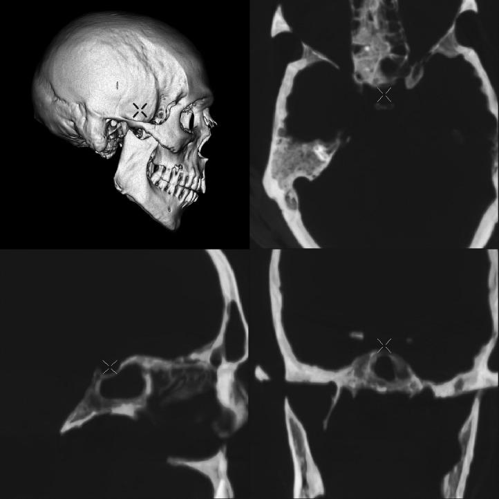
Materials and methods: Ten human skulls were x-rayed in conventional lateral cephalogram and then scanned with spiral CT. Twenty-eight linear and angular cephalometric measurements were registered on the 2D lateral cephalogram and compared to the same measurement on the 3D CT scan. Significance of the results was determined by t-test for paired differences (P < .05).
Results: No difference was found between 2D and 3D images for linear or ratio measurements. As for the angular cephalometric measurements, only the sella turcica dependent measurements, showed significant difference between 2D and 3D.
Conclusions: The compatibility of using most of the common orthodontic examined cephalometric measurements on 3D volume rendered image was proven except for the angular measurements that included sella anatomic landmark.
Figures
References
-
- Downs W. B. Variations in facial relationship—their significance in treatment and prognosis. Am J Orthod. 1948;34:812–840. - PubMed
-
- Downs W. B. The role of cephalometrics in orthodontic case analysis and diagnosis. Am J Orthod. 1952;38:162–182.
-
- Downs W. B. Analysis of dentofacial profile. Angle Orthod. 1956;26:191–212.
-
- Steiner C. C. The use of cephalometrics as an aid to planning and assessing orthodontic treatment. Am J Orthod. 1960;46:721–735.
-
- Ricketts R. M. Perspectives in the clinical application of cephalometrics. The first fifty years. Angle Orthod. 1981;51:115–150. - PubMed
Publication types
MeSH terms
LinkOut - more resources
Full Text Sources
Research Materials
