

Review
doi: 10.1186/1756-6606-3-28.
Molecular mechanisms of tiling and self-avoidance in neural development
Affiliations
- PMID: 20937126
- PMCID: PMC2959082
- DOI: 10.1186/1756-6606-3-28
Item in Clipboard
Review
Molecular mechanisms of tiling and self-avoidance in neural development
Mol Brain.
.
Abstract
Recent studies have begun to unravel the molecular basis of tiling and self-avoidance, two important cellular mechanisms that shape neuronal circuitry during development in both invertebrates and vertebrates. Dscams and Turtle (Tutl), two Ig superfamily proteins, have been shown to mediate contact-dependent homotypic interactions in tiling and self-avoidance. By contrast, the Activin pathway regulates axonal tiling in a contact-independent manner. These cell surface signals may directly or indirectly regulate the activity of the Tricornered kinase pathway and/or other intracellular signaling pathways to prevent the overlap between same-type neuronal arbors in the sensory or synaptic input field.
Figures
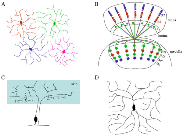
Examples of tiling and self-avoidance in vertebrates and invertebrates. A, A simplified diagram showing the tiling of vertebrate retinal ganglion neurons, based on results in [4]. B, Axonal tiling contributes to the organized columnar projection pattern of R7 and R8 photoreceptor neurons and L1 lamina neurons in the medulla of the Drosophila visual system. While L1 neurons arborize at both M1 and M5 sub-layers, R7 and R8 axons terminate at M6 and M3 sub-layers, respectively. Genetic dissection of neuronal circuit formation in the fly visual system has contributed significantly to our understanding of neuronal positioning, axon guidance and neuronal target selection (e.g. [53-57]). C, A schematic diagram showing the non-overlapping coverage of the receptive field by sister branches from a Pv mechanosensory neuron in leech, based on results in [14]. D, A simplified diagram showing self-avoidance in a Drosophila class IV da neuron, where sister branches tend to avoid each other.
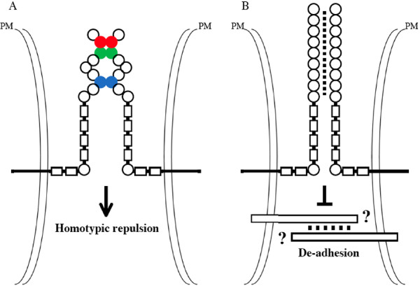
Proposed models for the action of Drosophila and vertebrate Dscams. A, Drosophila Dscam1 mediates homotypic repulsion in tiling and self-avoidance. The homophilic binding specificity of Dscam1 is determined by its Ig2 (red), Ig3 (green) and Ig7 (purple) domains. Half of Ig2, half of Ig3, and entire Ig7 domains are encoded by one of 12, 48, and 33 alternative exons, respectively. Thus, the extensive alternative splicing can generate 19,008 Dscam1 isoforms with different binding specificity. The binding between two Dscam isoforms only occurs if all three Ig domains (i.e. Ig2, Ig3 and Ig7) are identical. B, Vertebrate Dscams may mediate de-adhesion in tiling and self-avoidance by down-regulating the function of some unknown cell-type-specific adhesion molecules. The binding region on vertebrate Dscams is unknown, and presumably contains one or more of Ig domains. Circles, Ig domains. Rectangles, fibronectin type-III repeats. PM, plasma membrane
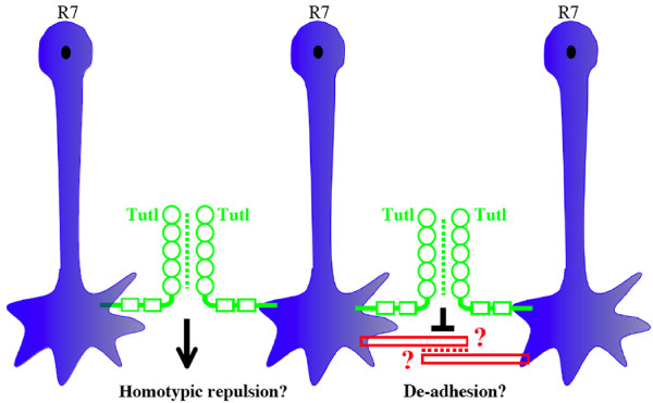
Proposed models for the action of Tutl. The homophilic binding between two Tutl proteins on opposing cell surface may be mediated by one or more of its Ig domains. Tutl may mediate homotypic repulsion. Alternatively, Tutl may mediate de-adhesion by antagonizing the function of certain R7-specific cell adhesion molecules. Circles, Ig domains; Rectangles, fibronectin type-III repeats.
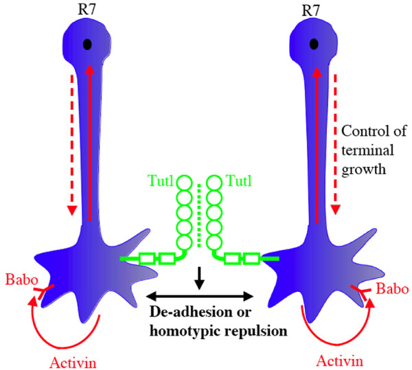
Co-operation between the Activin pathway and Tutl in the tiling of Drosophila R7 photoreceptor axons. Activin functions as an autocrine signal to activate its receptor Babo on R7 terminals, which in turn induces the phosphorylation of Smad2. The phosphorylated Smad2 is then transported by Importin α3 into the nucleus to regulate the expression of some unknown target genes, which directly control R7 terminal growth. This Activin-mediated intrinsic growth control functions together with Tutl-mediated homotypic interaction to regulate R7 tiling. Circles, Ig domains; Rectangles, fibronectin type-III repeats.
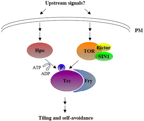
The function of the Trc signaling pathway in tiling and self-avoidance. Unidentified cell surface signals activate Hpo and TOR, which in turn up-regulate the activity of the Trc and Fry complex. Trc may then modulate the activity of certain cytoskeletal regulators to control the growth of neuronal arbors. PM, plasma membrane.
References
Publication types
MeSH terms
Substances
Grants and funding
LinkOut - more resources
Full Text Sources
