

Development of an in vitro alternative assay method for vaginal irritation
- PMID: 20937349
- PMCID: PMC3003762
- DOI: 10.1016/j.tox.2010.10.001
Development of an in vitro alternative assay method for vaginal irritation
Abstract
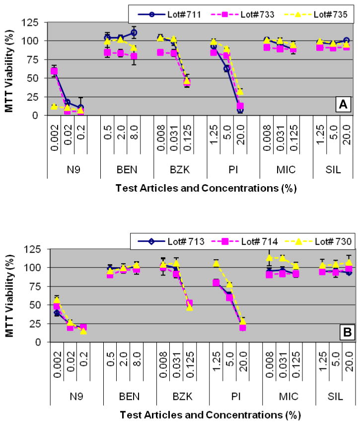
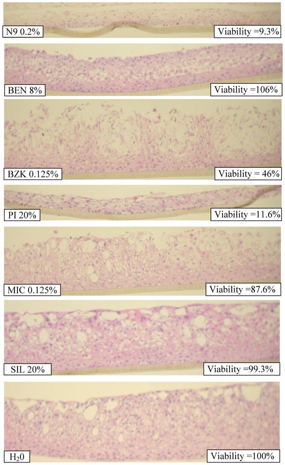
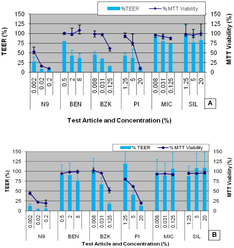
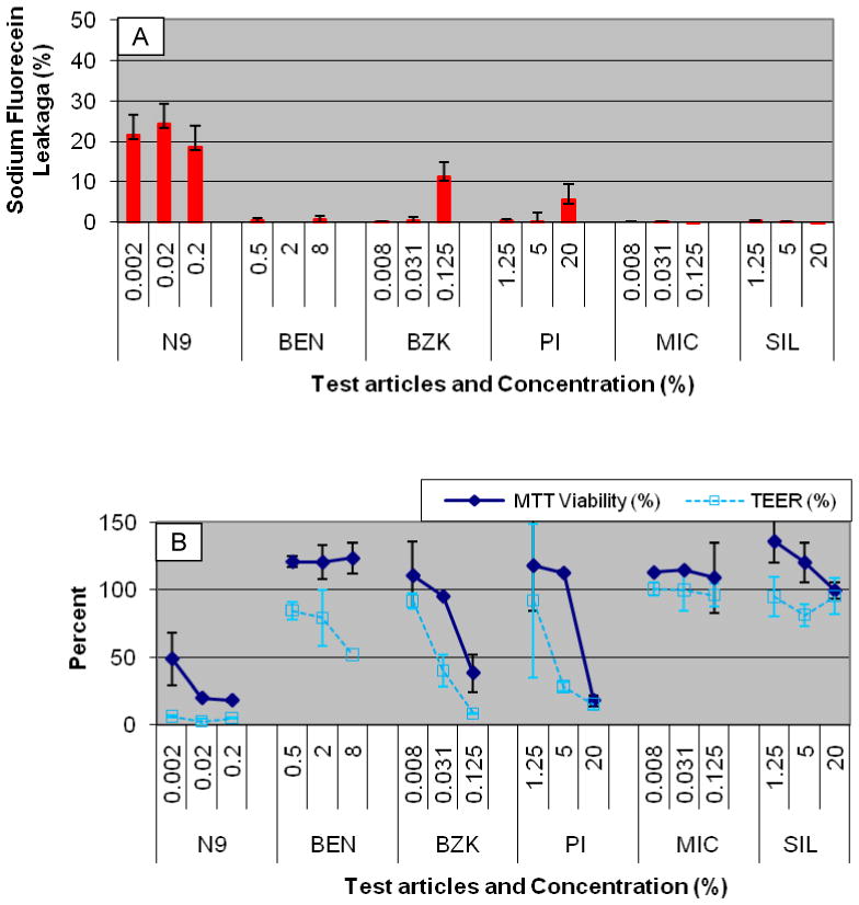
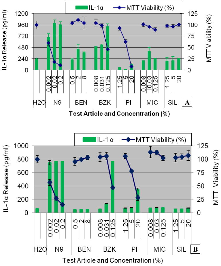
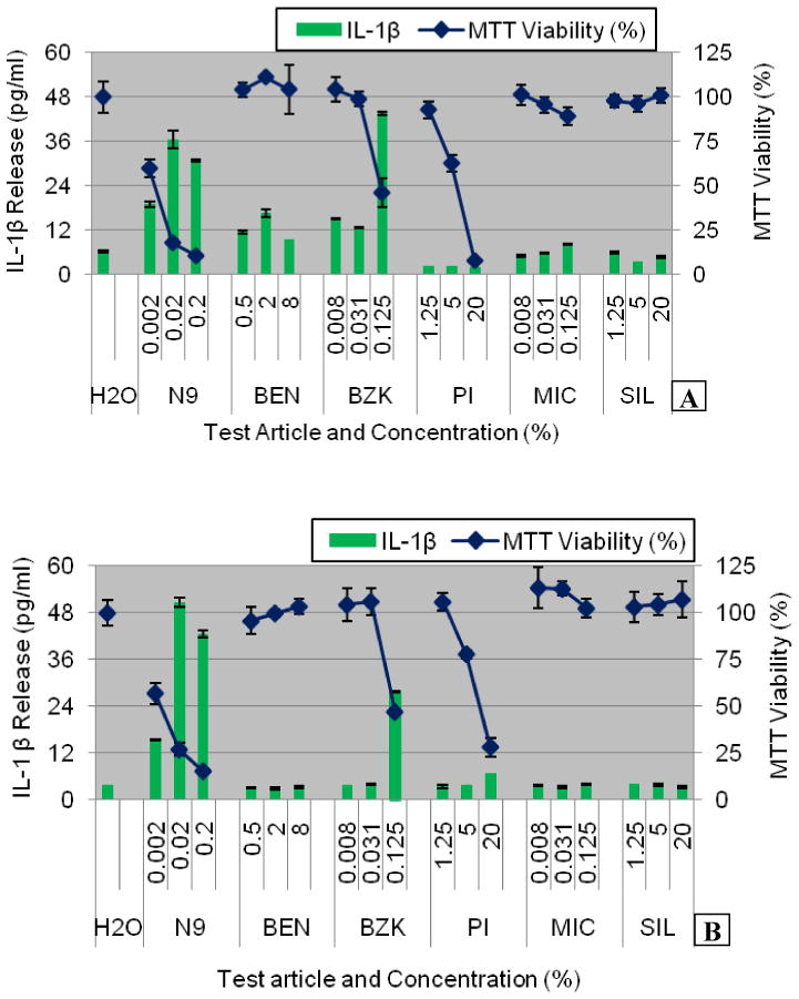
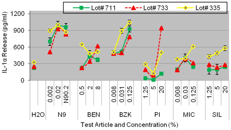
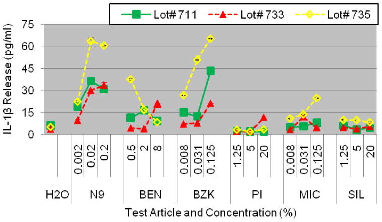
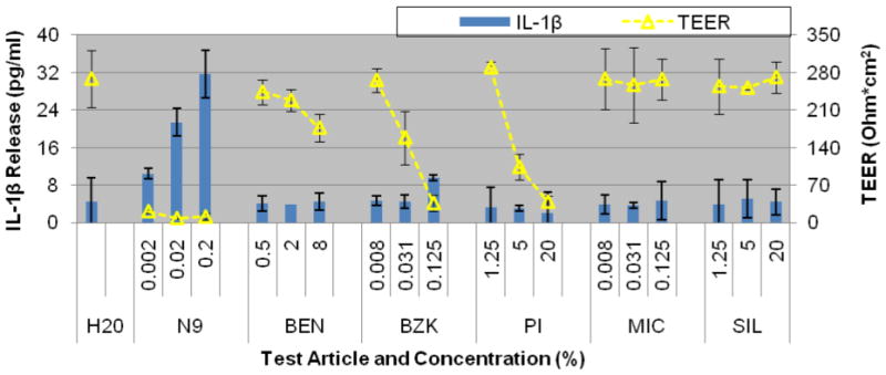
The vaginal mucosa is commonly exposed to chemicals and therapeutic agents that may result in irritation and/or inflammation. In addition to acute effects, vaginal irritation and inflammation can make women more susceptible to infections such as HIV-1 and herpes simplex virus 2 (HSV-2). Hence, the vaginal irritation potential of feminine care formulations and vaginally administered therapeutic agents is a significant public health concern. Traditionally, testing of such materials has been performed using the rabbit vaginal irritation (RVI) assay. In the current study, we investigated whether the organotypic, highly differentiated EpiVaginal™ tissue could be used as a non-animal alternative to the RVI test. The EpiVaginal tissue was exposed to a single application of ingredients commonly found in feminine hygiene products and the effects on tissue viability (MTT assay), barrier disruption (measured by transepithelial electrical resistance, TEER and sodium fluorescein (NaFl) leakage), and inflammatory cytokine release (interleukin (IL)-1α, IL-1β, IL-6, and IL-8) patterns were examined. When compared to untreated controls, two irritating ingredients, nonoxynol 9 and benzalkonium chloride, reduced tissue viability to <40% and TEER to <60% while increasing NaFl leakage by 11-24% and IL-1α and IL-1β release by >100%. Four other non-irritating materials had minimal effects on these parameters. Assay reproducibility was confirmed by testing the chemicals using three different tissue production lots and by using tissues reconstructed from cells obtained from three different donors. Coefficients of variation between tissue lots reconstructed with cells obtained from the same donor or lots reconstructed with cells obtained from different donors were less than 10% and 12%, respectively. In conclusion, decreases in tissue viability and barrier function and increases in IL-1α and IL-1β release appear to be useful endpoints for preclinical screening of topically applied chemicals and formulations for their vaginal irritation potential.
Copyright © 2010 Elsevier Ireland Ltd. All rights reserved.
Figures

References
-
- Achilles SL, et al. Microbicide efficacy and toxicity tests in a mouse model for vaginal transmission of Chlamydia trachomatis. Sex Transm Dis. 2002;29:655–664. - PubMed
-
- Ayehunie S, et al. Organotypic human vaginal-ectocervical tissue model for irritation studies of spermicides, microbicides, and feminine-care products. Toxicology in Vitro. 2006;20:689–698. - PubMed
-
- Bamforth SD, et al. Ultrastructural analysis of interleukin-1beta-induced leukocyte recruitment to the rat retina. Invest Ophthalmol & Visual Science. 1997;38:25–35. - PubMed
-
- Belec L, et al. Proinflammatory cytokine expression in cervicovaginal secretions of normal and HIV-infected women. Cytokine. 1995;7:568–74. - PubMed
Publication types
MeSH terms
Substances
Grants and funding
LinkOut - more resources
Full Text Sources
Other Literature Sources
