. 2010 Oct 18;191(2):259-67.
doi: 10.1083/jcb.201004150. Epub 2010 Oct 11.

Functional central spindle assembly requires de novo microtubule generation in the interchromosomal region during anaphase

Affiliations

Functional central spindle assembly requires de novo microtubule generation in the interchromosomal region during anaphase

Ryota Uehara et al. J Cell Biol. .

Abstract

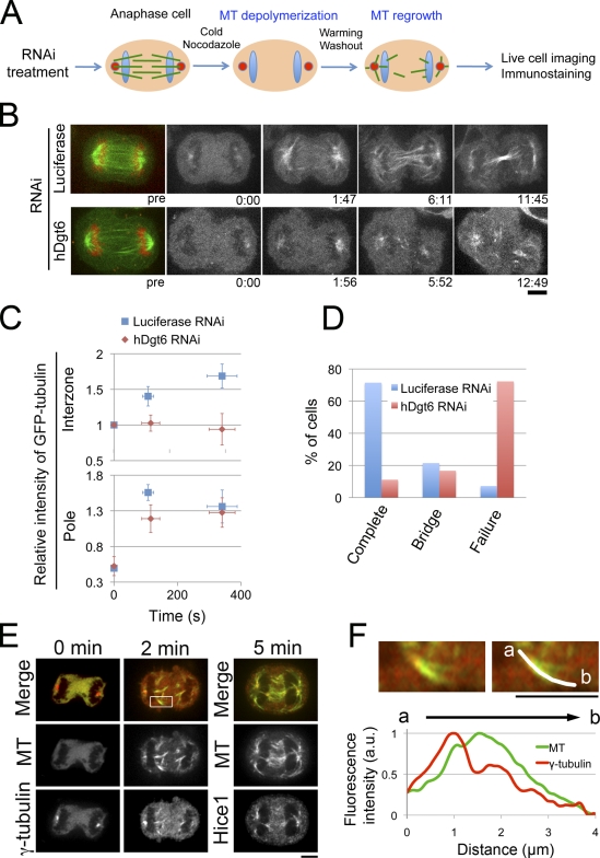
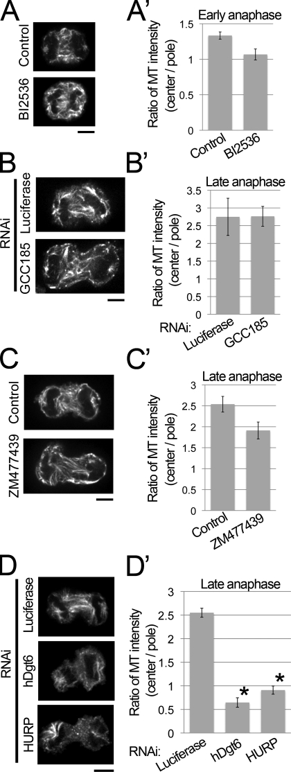
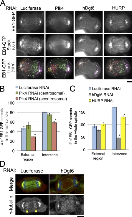
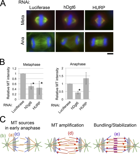
The central spindle forms between segregating chromosomes during anaphase and is required for cytokinesis. Although anaphase-specific bundling and stabilization of interpolar microtubules (MTs) contribute to formation of the central spindle, it remains largely unknown how these MTs are prepared. Using live imaging of MT plus ends and an MT depolymerization and regrowth assay, we show that de novo MT generation in the interchromosomal region during anaphase is important for central spindle formation in human cells. Generation of interchromosomal MTs and subsequent formation of the central spindle occur independently of preanaphase MTs or centrosomal MT nucleation but require augmin, a protein complex implicated in nucleation of noncentrosomal MTs during preanaphase. MTs generated in a hepatoma up-regulated protein (HURP)-dependent manner during anaphase also contribute to central spindle formation redundantly with preanaphase MTs. Based on these results, a new model for central spindle assembly is proposed.


Figures

Figure 1.
Figure 1.
Functional central spindle formation by augmin-dependent de novo MT generation in anaphase. (A) Schematic representation of the MT depolymerization/regrowth assay in anaphase cells. (B) GFP-tubulin–expressing cells treated with control (luciferase) or hDgt6 siRNA in the assay. The initial time point of observation is represented as 0:00 (min:s), which is ∼60 s after nocodazole was washed out. (C) Relative fluorescence intensity of GFP-tubulin in the interzone and the polar region of control (n = 14) and augmin-depleted cells (n = 18) in the assay. Means ± SEM (y axis) or SD (x axis) of single cells from six independent experiments are shown. (D) Completion or failure of cytokinesis after the assay in control (n = 14) and augmin-depleted cells (n = 18). Means of single cells from six independent experiments are shown. Bridge indicates the percentage of cells that possess an intercellular bridge at the end of the observation period. (E) Immunostaining of MT (green), γ-tubulin (red at 0 and 2 min), and Hice1 (red at 5 min) in anaphase cells during the MT regrowth phase. MT regrowth began at time 0. (F, top) A magnified image of the region indicated in the box in E. (bottom) MT and γ-tubulin intensities along an MT bundle. Chromosomes are located at the left. Bars, 5 µm.
Figure 2.
Figure 2.
HURP and augmin are required for de novo interzonal MT generation in the absence of preexisting MTs. (A–D) Immunostaining of MT in control, BI2536-treated (A), GCC185-depleted (B), ZM477439-treated (C), augmin- or HURP-depleted (D) cells fixed at 5 min after the MT depolymerization/regrowth treatment. Bars, 5 µm. (A’–D’) The ratio of central and polar MT intensities of anaphase cells in A–D. Means ± SEM of three independent experiments are shown (>16 cells were analyzed for each treatment). The ratio in both the augmin- and HURP-depleted cells but not in the other cells was significantly lower (augmin and HURP [asterisks], P < 0.001; other cells, P > 0.04; by t test) than that in the control cells.
Figure 3.
Figure 3.
Augmin, but not centrosomes or HURP, is critical for interzonal MT generation in unperturbed anaphase cells. (A) Single time frame images (top), 60-s stacked images (middle), and 10-s tracings (bottom) of EB1-GFP in RNAi-treated cells. Absence of the centrosome at the left pole of the monastral bipolar spindle of the Plk4-depleted cell was confirmed by z-axis confocal sectioning of the whole cell (Fig. S2 E). The following comets were distinguished: interzonal comets moving from left to right (acentrosomal to centrosomal side in Plk4-depleted cells; red), those moving in the opposite direction (green), comets moving in the external region on the left side (acentrosomal side in Plk-depleted cells; orange), or those on the opposite side (magenta). Light blue, interzonal comets with unknown directionalities; gray lines, borders between the external region and interzone. (B) Number of comets observed in each region of the half-spindle in control (blue) or Plk4-depleted cells (centrosomal or acentrosomal halves shown in green or red, respectively). Asterisks indicate statistically significant difference from the control (P < 0.008 by t test). The numbers of interzonal comets with unknown directionalities were 9.7 ± 2.3 (SEM) and 9.7 ± 3.5 in control and Plk4-depleted cells, respectively (not depicted). (C) The number of comets observed in each region of the whole spindle in control (blue), hDgt6-depleted (gray), and HURP-depleted cells (yellow) is shown. Means ± SEM of three independent experiments were shown (nine cells were analyzed for each region). Asterisks indicate statistically significant difference from control (P < 0.005 by t test). An identical dataset was used for luciferase RNAi cells in B and C. (D) Immunostaining of MTs (green), γ-tubulin (red), and chromosomes (blue) in control and hDgt6-depleted cells in anaphase. In the control cell, γ-tubulin was concentrated at both ends of the central spindle (arrowheads); however, such concentration was not observed in the augmin-depleted cell. Bars, 5 µm.
Figure 4.
Figure 4.
Physiological importance of de novo interzonal MT generation in anaphase. (A) Immunostaining of MT (green), γ-tubulin (red), and chromosomes (blue) in control, augmin-depleted, or HURP-depleted cells in metaphase (meta) or anaphase (ana). Bar, 5 µm. (B) MT intensities in the metaphase spindle or interzone in anaphase. Means ± SD of three independent experiments are shown (>18 cells were analyzed for each treatment). Depletion of hDgt6 and HURP resulted in significant reduction in MT amounts in metaphase (*, P < 0.01), whereas only the former caused significant reduction in anaphase interzonal MTs (*, P < 0.0003 for hDgt6-depleted anaphase cells; P > 0.09 for HURP-depleted anaphase cells). (C) A model for the central spindle formation process. In anaphase, MTs that are formed in preanaphase (a; orange), those constantly nucleated from centrosomes (b; green), and those nucleated in the interzone near the chromosomes in an HURP-dependent manner (c; blue) are available as MT sources. (d; red) These MTs are used as the templates for MT amplification mediated by augmin and γ-TuRC. (e; purple) All of the interzonal MTs formed by processes a–d are stabilized and bundled by antiparallel MT-bundling proteins, resulting in robust central spindle formation.

References

    1. Alsop G.B., Zhang D. 2003. Microtubules are the only structural constituent of the spindle apparatus required for induction of cell cleavage. J. Cell Biol. 162:383–390 10.1083/jcb.200301073 - DOI - PMC - PubMed
    1. Altan-Bonnet N., Sougrat R., Liu W., Snapp E.L., Ward T., Lippincott-Schwartz J. 2006. Golgi inheritance in mammalian cells is mediated through endoplasmic reticulum export activities. Mol. Biol. Cell. 17:990–1005 10.1091/mbc.E05-02-0155 - DOI - PMC - PubMed
    1. Bastiaens P., Caudron M., Niethammer P., Karsenti E. 2006. Gradients in the self-organization of the mitotic spindle. Trends Cell Biol. 16:125–134 10.1016/j.tcb.2006.01.005 - DOI - PubMed
    1. Belmont L.D., Hyman A.A., Sawin K.E., Mitchison T.J. 1990. Real-time visualization of cell cycle-dependent changes in microtubule dynamics in cytoplasmic extracts. Cell. 62:579–589 10.1016/0092-8674(90)90022-7 - DOI - PubMed
    1. Bettencourt-Dias M., Rodrigues-Martins A., Carpenter L., Riparbelli M., Lehmann L., Gatt M.K., Carmo N., Balloux F., Callaini G., Glover D.M. 2005. SAK/PLK4 is required for centriole duplication and flagella development. Curr. Biol. 15:2199–2207 10.1016/j.cub.2005.11.042 - DOI - PubMed

Publication types