. 2010 Oct 18;191(2):367-81.
doi: 10.1083/jcb.201008051. Epub 2010 Oct 11.

Family-wide characterization of the DENN domain Rab GDP-GTP exchange factors

Affiliations

Family-wide characterization of the DENN domain Rab GDP-GTP exchange factors

Shin-ichiro Yoshimura et al. J Cell Biol. .

Abstract

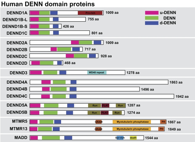
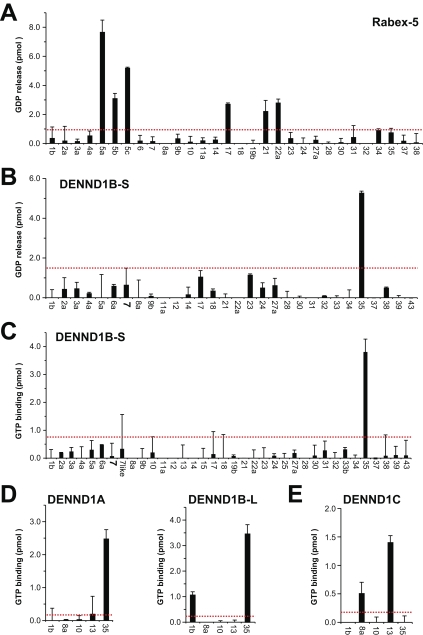
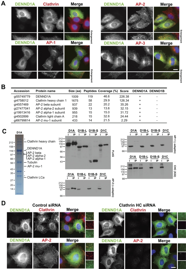
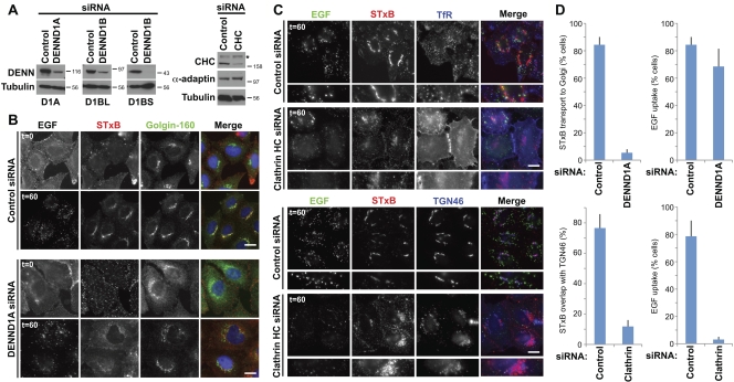
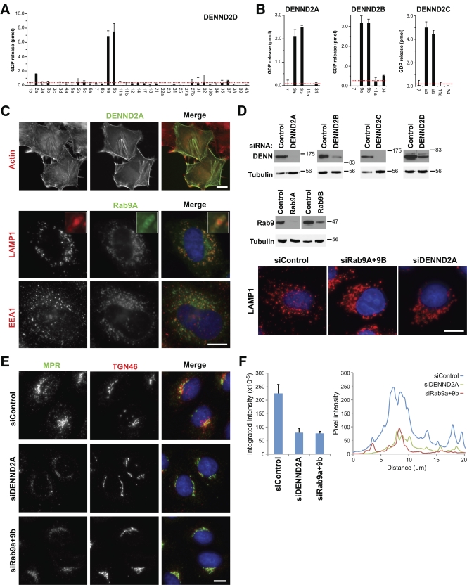
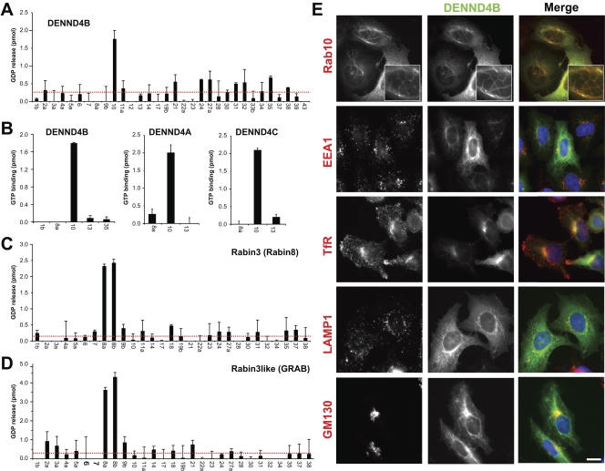
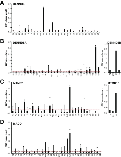
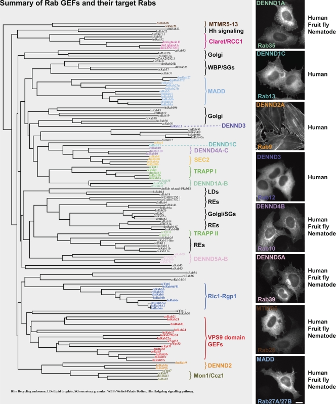
A key requirement for Rab function in membrane trafficking is site-specific activation by GDP-GTP exchange factors (GEFs), but the majority of the 63 human Rabs have no known GEF. We have performed a systematic characterization of the 17 human DENN domain proteins and demonstrated that they are specific GEFs for 10 Rabs. DENND1A/1B localize to clathrin patches at the plasma membrane and activate Rab35 in an endocytic pathway trafficking Shiga toxin to the trans-Golgi network. DENND2 GEFs target to actin filaments and control Rab9-dependent trafficking of mannose-6-phosphate receptor to lysosomes. DENND4 GEFs target to a tubular membrane compartment adjacent to the Golgi, where they activate Rab10, which suggests a function in basolateral polarized sorting in epithelial cells that compliments the non-DENN GEF Sec2 acting on Rab8 in apical sorting. DENND1C, DENND3, DENND5A/5B, MTMR5/13, and MADD activate Rab13, Rab12, Rab39, Rab28, and Rab27A/27B, respectively. Together, these findings provide a basis for future studies on Rab regulation and function.


Figures

Figure 1.
Figure 1.
DENN proteins form a large family in human cells. A schematic showing the human DENN domain proteins, with the upstream (u-DENN), core DENN, and downstream (d-DENN) regions indicated. Additional domains likely to be of relevance for DENN targeting or regulation are marked and color coded. Sequence alignments of DENNs were done with ClustalX (Chenna et al., 2003) or MUSCLE (Edgar, 2004), and the results were visualized and manipulated with Jalview (Waterhouse et al., 2009). Linear sequence motifs were browsed in the ELM database (Gould et al., 2010). Accession numbers used for this analysis are listed in Table S1.
Figure 2.
Figure 2.
DENND1A/1B are GEFs for Rab35. (A) Human Rabex-5 was tested against a representative panel of human Rab proteins using the GDP-releasing assay. In brief, 10 µg of each GST-tagged Rab to be tested was incubated in 50 mM Hepes-NaOH, pH 6.8, 0.1 mg/ml BSA, 125 µM EDTA, 10 µM Mg-GDP, and 5 µCi [3H]-GDP (10 mCi/ml; 5,000 Ci/mmol) in a total volume of 200 µl for 15 min at 30°C to load the Rab with the radioactive GDP probe. For standard GDP-releasing GEF assays, 100 µl of the loading reaction was then mixed with 10 µl of 10 mM Mg-GTP and 10 nM His6-tagged Rabex-5 purified from bacteria or a buffer control, then adjusted to 120 µl final volume with assay buffer. The GEF reaction occurred for 20 min at 30°C. After this, 2.5 µl was taken for a specific activity measurement; the remainder was split into two tubes, then incubated with 500 µl of ice-cold assay buffer containing 1 mM MgCl2 and 20 µl of packed glutathione-sepharose for 60 min at 4°C to separate Rab–GDP complexes from free “released” GDP. After washing three times with 500 µl of ice-cold assay buffer, the sepharose was transferred to a vial containing 4 ml of scintillation fluid and counted. The amount of nucleotide exchange was calculated in pmoles of GDP released. (B and C) A representative panel of human Rab proteins was tested against 10 nM of His6-tagged DENND1B-S in the GDP-releasing (B) or GTP-binding assay (C). For GTP-binding assays, the following modifications were made: only unlabeled GDP was used in the loading reaction; in the GEF reaction, 0.5 µl of 10 mM GTP and 1 µCi [35S]-GTPγS (10 mCi/ml; 5000 Ci/mmol) were used. The amount of nucleotide exchange was calculated in pmoles of GTP bound. (D and E) Human DENND1A (D), DENND1B-L (D), and DENND1C (E) were tested against a subset of Rab35-related Rabs using the GTP-binding assay. For these assays, 10 nM of FLAG-tagged DENND1A or DENND1C purified from HeLa cells, or 10 nM of His6-tagged DENND1B-L purified from bacteria were used. Errors bars show the standard error from the mean. The red line marks double the median value taken as a threshold.
Figure 3.
Figure 3.
Localization of DENND1A is clathrin dependent. (A) HeLa cells expressing EGFP-tagged DENND1A (green) were fixed and then stained with antibodies to clathrin heavy chain, and the AP-1, AP-2, and AP-3 clathrin adaptor complexes (red). DNA was stained with DAPI (blue). (B) DENND1A and DENND1B complexes were analyzed by mass spectrometry. The proteins scored highest by the Sequest search algorithm are listed in the table. (C) HeLa cells were transfected with constructs encoding FLAG-tagged DENND1A, DENND1B-L, DENND1B-S, and DENND1C for 48 h. The cells were washed from the dish using PBS with 1 mM EDTA, and the cell pellets were lysed for 20 min on ice in 1 ml cell of lysis buffer (50 mM Tris-HCl, pH 7.4, 1 mM EDTA, 150 mM NaCl, 0.5% Triton X-100, and protease inhibitors cocktails). The FLAG-tagged proteins were immunoprecipitated from the clarified lysate using 20 µl of anti-FLAG M2 affinity gel (Sigma-Aldrich) for 4 h at 4°C. The pellet was washed three times in 1 ml of cell lysis buffer, and bound proteins were eluted with 1 ml of 200 µg/ml FLAG peptide in TBS and then precipitated for 60 min on ice using 10% trichloroacetic acid. The FLAG-tagged DENND1A, DENND1B-L, DENND1B-S, and DENND1C complexes were analyzed by SDS-PAGE on 4–12% gradient gels and Coomassie blue staining, or Western blotted for clathrin heavy chain (CHC) and the AP-1 and AP-2 clathrin adaptors on 10% gels. Asterisks mark proteins that nonspecifically bind to FLAG-agarose and were found in negative control conditions. Molecular mass standards are indicated in kilodaltons. (D) HeLa cells expressing EGFP-tagged DENND1A (green) were transfected for 72 h with siRNA duplexes targeting the clathrin heavy chain, fixed, and then stained with antibodies for clathrin and the AP-2 clathrin adaptor (red). DNA was stained with DAPI (blue). Enlargements are shown to the right to more clearly demonstrate the overlap between DENND1A (green), and clathrin or AP2 (red) in control cells, and the loss of punctate DENND1 staining after clathrin heavy chain depletion. Bars, 10 µm.
Figure 4.
Figure 4.
DENND1A is required for Rab35-dependent Shiga toxin trafficking to the trans-Golgi network. (A) HeLa cells expressing EGFP-tagged DENND1A and the long or short forms of DENND1B were transfected with siRNA duplexes to DENND1A, DENND1B, clathrin heavy chain (CHC), or a nonspecific control for 72 h, then Western blotted as indicated. Molecular mass standards are indicated in kilodaltons. The asterisk indicates a nonspecific cross reaction of the clathrin heavy chain antibody. (B) Dual EGF and STxB uptake assays were performed for 60 min as described previously (Fuchs et al., 2007) in cells transfected with control or DENND1A duplexes for 72 h. Cells were fixed and then stained for the Golgi marker golgin-160. (C) Uptake assays were performed as in B using cells transfected with CHC siRNA duplexes. Cells were fixed and then stained for the transferrin receptor (TfR) to mark recycling endosomes or the TGN marker TGN46. Bars, 10 µm. (D) The extent of EGF and Shiga toxin uptake under the various conditions was measured and is plotted in the graphs (n = 3). ImageJ was used to measure colocalization of markers. Error bars indicate standard error of the mean.
Figure 5.
Figure 5.
The DENND2 family regulates Rab9 and lysosomes. (A) A representative panel of human Rab proteins was tested against 10 nM of His6-tagged human DENND2D purified from bacteria using the GDP-releasing assay. (B) Human DENND2A, DENND2B, and DENND2C were expressed as His6-tagged protein in bacteria and then tested against a subset of Rab9-related Rabs. Errors bars show the standard error of the mean. The red line marks double the median value. (C) HeLa cells were transfected with EGFP-tagged DENND2A or Rab9A (green), fixed after 24 h, and stained with the antibodies indicated (red). DNA was stained with DAPI (blue). Inset enlargements are shown to more clearly demonstrate the relationship between Rab9 (green) and LAMP1 (red), which suggests that Rab9 is present on the lysosome membrane. (D) HeLa cells expressing EGFP-tagged Rab9 or DENND2 constructs as indicated were transfected with control, Rab9A, Rab9B, and DENND2A-D siRNA duplexes for 72 h. Western blotting with EGFP antibodies confirmed depletion of the target proteins, whereas tubulin showed that loading was equal for all samples. HeLa cells transfected with control, Rab9A and Rab9B, and DENND2A-D siRNA duplexes for 72 h were fixed, then stained for LAMP1 (red) and DAPI to detect DNA (blue). Molecular mass standards are indicated in kilodaltons. (E) HeLa cells transfected with control, Rab9A and Rab9B, and DENND2A siRNA duplexes for 72 h were fixed, then stained for MPR (green) and TGN46 (red). DNA was stained with DAPI (blue). Bars, 10 µm. (F) Fluorescence intensity for MPR staining from E was measured using ImageJ by drawing a box around the entire cell area and integrating the total signal. An equivalent area with no cell was subtracted for the background. This was performed for 24 cells, and the mean and standard error are plotted on the bar graph. A 20 × 1 µm line measurement was performed across the nuclear region where MPR staining is most clustered. The pixel intensity along the line is plotted in the graph for control, DENND2A, and Rab9a- and Rab9b-depleted cells.
Figure 6.
Figure 6.
DENND4 family proteins are specific GEFs for Rab10. (A) A representative panel of human Rab proteins was tested against 10 nM of human DENND4B expressed as a FLAG-tagged protein in HeLa cells using the GDP-releasing assay. (B) Human DENND4A, DENND4B, and DENND4C were tested against a subset of Rab10-related Rabs using the GTP-binding assay. Again, 10 nM of each FLAG-tagged DENN protein purified from HeLa cells was used for these assays. Error bars show the standard error of the mean. The red line marks double the median value. (C and D) A representative panel of human Rab proteins was tested against 10 nM of His6-tagged human Rabin3/Rabin8 (C) and Rabin3-like/GRAB purified from bacteria using the GDP-releasing assay (D). Error bars indicate standard error of the mean. (E) HeLa cells expressing EGFP-tagged DENND4B were transfected with mCherry-tagged Rab10, or stained for the markers indicated. 4× enlargements of the Rab10-positive tubules are shown in the top panels. Bar, 10 µm.
Figure 7.
Figure 7.
Family-wide assignment of DENN specificity. Human DENND3 (A), DENND5A and DENND5B (B), MTMR5 and MTMR13 (C), and MADD (D) were tested against a representative panel of human Rab proteins using the GDP-releasing assay. All assays used 10 nM of FLAG-tagged DENN protein purified from HeLa cells. Errors bars show the standard error of the mean. The red line marks double the median value.
Figure 8.
Figure 8.
A summary of Rab GEFs indicating their target Rabs. Human (Homo sapiens, hs), fruit fly (Drosophila melanogaster, dm), and nematode (C. elegans, ce) Rabs and budding yeast (Saccharomyces cerevisiae, sc) Ypts were aligned using ClustalX and plotted using NJplot (Larkin et al., 2007). The alignment is annotated to show the known Rab GEFs: TRAPP, Sec2, the Vps9 domain family, Ric1-Rgp1, Mon1-Ccz1, claret, and the DENN domain family. Images to the right indicate the typical localization of the DENN domain family in HeLa cells. The pattern of conservation is summarized in the text to the right. The accession nos. for human, mouse, zebrafish, fruit fly, and nematode DENN domains proteins are listed in Table S1. Bar, 10 µm.

References

    1. Allaire P.D., Marat A.L., Dall’Armi C., Di Paolo G., McPherson P.S., Ritter B. 2010. The Connecdenn DENN domain: a GEF for Rab35 mediating cargo-specific exit from early endosomes. Mol. Cell. 37:370–382 10.1016/j.molcel.2009.12.037 - DOI - PMC - PubMed
    1. Azzedine H., Bolino A., Taïeb T., Birouk N., Di Duca M., Bouhouche A., Benamou S., Mrabet A., Hammadouche T., Chkili T., et al. 2003. Mutations in MTMR13, a new pseudophosphatase homologue of MTMR2 and Sbf1, in two families with an autosomal recessive demyelinating form of Charcot-Marie-Tooth disease associated with early-onset glaucoma. Am. J. Hum. Genet. 72:1141–1153 10.1086/375034 - DOI - PMC - PubMed
    1. Babbey C.M., Ahktar N., Wang E., Chen C.C., Grant B.D., Dunn K.W. 2006. Rab10 regulates membrane transport through early endosomes of polarized Madin-Darby canine kidney cells. Mol. Biol. Cell. 17:3156–3175 10.1091/mbc.E05-08-0799 - DOI - PMC - PubMed
    1. Barr F., Lambright D.G. 2010. Rab GEFs and GAPs. Curr. Opin. Cell Biol. 22:461–470 10.1016/j.ceb.2010.04.007 - DOI - PMC - PubMed
    1. Behnia R., Munro S. 2005. Organelle identity and the signposts for membrane traffic. Nature. 438:597–604 10.1038/nature04397 - DOI - PubMed

Publication types

MeSH terms

Substances